


Cataract Overview
Cataract Overview
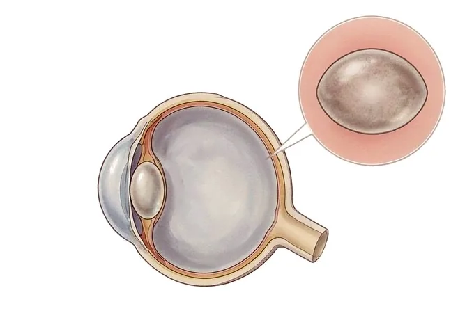
Ageing changes the lens of the eye the way it changes a window: not by breaking it, but by making it less clear. The lens sits behind the pupil and does the focusing, bending light onto the retina at the back of the eye, and it is built from proteins laid down in precise layers. Over decades those proteins gather and lose their order, and layers that once let light pass cleanly begin to scatter it. That is a cataract, and the most useful thing to hold on to about it is what the word does not mean. It is not a growth, not a film laid over the eye, and not a disease of the eye as a whole. It is one part of the eye changing, in a way that can be corrected in an unusually direct fashion: the clouded lens is removed and a clear artificial one is put in its place. Three consequences follow. It is common, because it is part of ageing rather than an affliction that arrives from outside. It is gradual, because the change in the lens takes years. And the result of correcting it is not automatic, because the eye the new lens focuses into matters at least as much as the lens that has been taken out. That is why a cataract assessment spends as much time on the retina and the optic nerve as it does on the lens, and why the measurements taken before surgery are of the whole eye and not only of the opacity in it.
-
A lens, not the whole eye
the operation replaces a clouded lens; the retina and the optic nerve still decide the outcome
-
Waiting for it to ripen is out of date
timing is set by what it costs you in daily life, not by how clouded it looks
-
Glare is a separate measure
light scatter can end night driving while the acuity chart still reads well
-
48 hrs
from complete records to a written assessment plan
What a cataract is, and what it is not
A cataract is a clouding of the lens inside the eye. The lens is a small, transparent structure behind the pupil, about the size of a lentil, and it does the focusing: it bends incoming light so that it lands as a sharp image on the retina at the back of the eye. It is built from proteins arranged in layers with an almost crystalline order, and that order is what makes it transparent.
The most useful thing to understand about the word is what it does not mean. A cataract is a change in one part of the eye. It is not a disease of the eye.
- It is not a growth, and not a film over the eye. Nothing is added to the eye. The lens that is already there loses its clarity from within, which is why no drops, exercises or diet dissolve it.
- It does not usually mean the eye is otherwise damaged. Most eyes with a cataract are healthy apart from the lens, and once the lens is replaced they see well. A minority are not, and the assessment exists to find that out beforehand.
- It does mean the outcome is a shared result. The operation replaces the lens; the retina, the optic nerve and the cornea receive the image the new lens forms. Where those are healthy, the result is usually good. Where they are not, the lens is still worth replacing — but what it can be expected to deliver is different, and saying so in advance is part of the assessment.
Why it happens at all. Lens proteins are laid down before birth and are never replaced. Over a lifetime they are exposed to light, to oxygen and to the by-products of metabolism, and they gradually cross-link and clump. The clumps scatter light instead of transmitting it. Nothing about this process requires an external cause, which is why an ophthalmologist asked what caused a cataract in a person in their seventies will generally answer that time did.
What has changed is not the eye but who reaches surgery. Cataract surgery is the most frequently performed operation in medicine, and the reason is not that cataracts have become more common but that the operation has become short, safe and predictable enough to offer early. The threshold has moved from a lens that is visibly white and blocking vision to a lens that is making night driving, reading or working uncomfortable — a change in practice that most patients have not been told about.
The four places a cataract forms
Cataracts are named by where in the lens the clouding sits, and the position explains the symptoms better than the size of the opacity does. Four patterns cover almost everything seen in clinic.
| What it is called | Where the clouding sits | What it typically does to sight |
|---|---|---|
| Nuclear cataract | In the centre of the lens, the part that does most of the focusing | Distance vision blurs gradually and colours yellow; near vision may briefly improve, as the denser centre acts like a stronger lens |
| Cortical cataract | In the outer layers, as wedge-shaped spokes running from the edge inwards | Glare and scattered light, with vision that varies with the lighting and can be better in dim rooms than in bright ones |
| Posterior subcapsular cataract | At the very back of the lens, on the visual axis itself | Reading and near vision affected early, with marked glare and halos; often linked to steroid use or diabetes, and can progress quickly |
| Congenital or developmental cataract | Present from birth or forming in childhood, sometimes partial and sometimes dense | Depends entirely on how much of the axis it covers; a dense one affecting one eye has to be dealt with early to protect the development of vision |
| Secondary cataract | Forming in a lens affected by something else, rather than by age alone | Follows eye injury, uveitis, previous eye surgery, long-term steroids or diabetes; tends to behave like the condition that caused it |
Why the label matters more than the size. Two cataracts of the same measured density behave quite differently depending on where in the lens they sit. A small posterior subcapsular opacity sitting on the visual axis can trouble reading and night driving more than a much larger nuclear one, because of where it is rather than how big it is. That is why the slit lamp description in the notes is worth reading, and why a patient whose acuity chart looks reasonable can be describing a genuine and disabling problem.
One term worth knowing. A “secondary cataract” is sometimes used by patients to describe a re-clouding months or years after surgery. What happens in that situation is different: the artificial lens stays clear, and a membrane behind it thickens. It is treated with a brief laser procedure rather than with another operation — a distinction worth making, because the two sound identical and are not.
What moves a cataract up or down the list
A cataract is not an emergency, and most can be watched. What sorts them into those that can wait and those that should be dealt with is a short list of features, and each is checked at the same assessment.
| Feature | Why it changes the plan |
|---|---|
| What it is costing you in daily life | The decisive question, and not a measurement. Difficulty driving at night, reading, working, or recognising faces is reason enough, whatever the chart says |
| Glare out of proportion to the acuity | Light scatter is a separate problem from blurring and can make night driving unsafe while measured vision still looks satisfactory |
| The view to the back of the eye being blocked | A dense lens prevents proper examination and monitoring of the retina and the optic nerve, which matters in diabetic eye disease, glaucoma and macular disease |
| The state of the other eye | Good vision in one eye can compensate for a poor one for years; if the good eye is itself at risk, the cataract becomes more urgent |
Where the old rule about waiting fits in. The advice to wait until a cataract has “ripened” belongs to an era when surgery required a large incision and a dense, hard lens was easier to remove whole. Neither is true of modern surgery. A very advanced cataract is now the harder one to operate on: the lens is harder, the ultrasound energy needed is greater, and the operation takes longer, while the recovery is no better than it would have been earlier. The timing question is therefore about the patient, not about the lens.
How a cataract is assessed
The assessment is short and is designed to answer three questions in a single pass: how much the lens is costing you, whether the rest of the eye is healthy, and what power of replacement lens the eye would need.
| Step | The question it answers |
|---|---|
| Vision and refraction | How much is the lens costing you, and would a change of glasses help in the meantime — a genuine answer while a cataract is early |
| Slit lamp and dilated examination | What sort of cataract is it, how far has it advanced, and is the retina, optic nerve and cornea behind it healthy? |
| Measurements for surgery | What power of lens does this particular eye need — calculated from its length and the curvature of its cornea before the operation, not during it |
Why the three are read together. Each step can mislead on its own. A good acuity reading can coexist with disabling glare; a cataract that looks straightforward can sit in an eye with macular disease that changes what can be promised; a refraction that improves things can hide the fact that the lens, not the prescription, is the limiting factor. Where the three agree, the plan is straightforward. Where they disagree, the disagreement is the finding that decides whether to operate, when, and what to say about the likely result.
What patients want is usually an answer, not a sequence — but the sequence is what makes the answer trustworthy. The whole design of the assessment is that nobody is offered an operation on the strength of a cloudy lens alone, and nobody is told to wait on the strength of a good chart alone.
Why patients travel to China for cataract surgery
A cataract is a mechanical problem with a mechanical solution, and what makes the international pathway worth considering is a large surgical volume, short waits, and an assessment that is genuinely complete before the decision is made.
The assessment arrives complete, not in instalments. Vision, refraction, slit lamp examination, dilated examination of the retina, measurement of the eye and the calculation of the replacement lens power are organised within a single facility, so a question that in many health systems spans several appointments and several waiting lists is answered inside one short visit, with the measurements taken while the patient is still there.
Previous imaging and records are reviewed before arrival. Earlier prescriptions, retinal imaging, optical coherence tomography and any previous operation notes are read in advance — the images themselves, not only the summary — so that the first consultation starts from what is already known rather than rediscovering it.
Where the eye is complex, the plan is made by a team. Retina, glaucoma, cornea and cataract specialists review the same eye together where diabetic retinopathy, macular disease, glaucoma or previous refractive surgery are involved, rather than each seeing the patient separately. Where a dense cataract prevents the retina from being seen, that is established before surgery and planned for, not discovered afterwards.
Both eyes are planned as one pathway, two decisions. The second eye is assessed on its own measurements, and the interval between operations is set by the healing of the first. Where lens choice interacts with what the other eye will need, that is discussed before the first operation rather than after it.
Costs are set out before commitment. An itemised estimate covering the assessment, the operation, the lens chosen and follow-up is provided in advance; international-desk staff coordinate with the patient and with any insurer or assistance company throughout, and written summaries, surgical notes and follow-up plans are issued in English for the doctor at home.










