


Brain Metastases Overview
Brain Metastases Overview
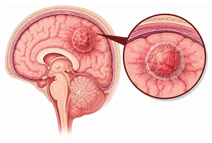
A brain metastasis is not a new cancer. It is a deposit of the cancer that already exists somewhere else in the body, carried to the brain through the bloodstream, and growing where it lands. That single fact explains why this condition is treated differently from a tumour that begins in the brain: the disease being managed is the cancer as a whole, and the deposits in the brain are the part of it that happens to threaten the functions the brain performs.
-
Commonest
the most frequent tumours found inside the skull in adults — more common than tumours that begin in the brain
-
Number and site
how many deposits there are and where they sit, which decides whether focused treatment is possible
-
Markers
the molecular profile of the cancer, which can make a targeted drug the most effective route rather than surgery
-
48 hrs
from complete records to a personalized MDT plan
What a brain metastasis is
Cancer spreads when cells leave the place where the tumour began, travel through the blood or the lymph channels, and settle somewhere else. When that destination is the brain, the resulting deposit is called a brain metastasis. It is the same disease as the original cancer — a lung cancer that has reached the brain is still lung cancer, and is treated with the drugs that work against lung cancer — and this is why the first question in planning treatment is not "what is in the brain" but "what is this cancer, and what is it sensitive to".
Two differences follow from the site. The first is space. The skull is a closed box, so a deposit that grows takes up room that the brain needs, and the swelling that surrounds it adds to the pressure. That is why symptoms can appear from a deposit that is still small, and why reducing that swelling — usually with a steroid — can bring relief within days even before the deposit itself is treated.
The second is function. A deposit sitting in the part of the brain that controls movement on one side, or in the area that produces speech, or in the pathways that carry signals down to the limbs, causes disability that is specific to where it is, not to how large it is. A deposit in the back of the brain can block the flow of fluid and cause pressure that way. And because these deposits are distributed by the bloodstream rather than growing from one point, they are often multiple, which is the single most important fact in deciding how they are treated.
What does not follow is that spread to the brain means nothing further can be done. The range of treatments has widened considerably, and the order in which they are used depends on findings that are made at the outset. For that reason the useful thing to establish early is a complete picture: what the cancer is, what it carries molecularly, how many deposits there are, how the disease elsewhere is behaving, and what the patient is still able to do.
Which cancers spread to the brain
Any cancer can spread to the brain, but a small number do so often enough that the possibility is monitored for. By frequency, the ones that matter most are:
- Lung cancer — the commonest source by a wide margin, and the one in which a deposit is most often found on a staging scan before it causes symptoms. It is also the type in which molecular testing most often changes the treatment, because several targeted drugs reach the brain effectively.
- Breast cancer — particularly the HER2-positive and triple-negative subtypes, and often later in the course of the disease. In HER2-positive disease the brain has become a more frequent site precisely because control elsewhere has improved.
- Melanoma — a cancer with a well-recognised tendency to spread to the brain, sometimes with several deposits at once and sometimes with bleeding into them. It is also the type in which immunotherapy has altered what is possible.
- Colorectal cancer — less common than the three above, and usually in the setting of disease that has already spread to the liver or lungs.
- Kidney cancer — occasionally presenting with a single deposit that bleeds, and one of the types in which surgical removal is considered more readily.
- Other types and unknown primary — cancers of the ovary, the uterus, the head and neck and others may spread to the brain, and in a minority of patients a deposit is found before the original cancer has been identified. In that situation finding the source, by imaging and sometimes by biopsy, becomes part of the treatment planning rather than an academic question.
The type matters for a practical reason. Each of these cancers carries its own set of molecular markers, and those markers determine whether a drug that works well inside the brain is available. A deposit from a lung cancer with an EGFR mutation, a breast cancer that is HER2-positive, or a melanoma with a BRAF mutation are all situations in which drug treatment given first can be the most effective approach — a point that is easy to miss if the brain deposit is considered in isolation.
How the extent is assessed
There is no stage number for a brain metastasis. What replaces it is a set of findings, and each of them changes what is offered. These are the ones that are recorded:
| Finding | What it means | How it is used |
|---|---|---|
| Number of deposits | Whether there is one, a few, or many — counted on contrast-enhanced MRI rather than on CT | Few deposits can be treated with focused, precisely aimed radiotherapy; many deposits usually need whole-brain treatment |
| Size and site | How large each deposit is, and whether it sits in an area that controls movement, speech or vision, or blocks the flow of fluid | Determines whether an operation is helpful, and whether the radiotherapy can be delivered safely around critical tissue |
| Fluid-space involvement | Whether cancer cells have reached the cerebrospinal fluid that surrounds the brain and spinal cord | Changes the treatment entirely, and is looked for specifically when there are symptoms beyond the deposits themselves |
| Disease outside the brain | How much cancer there is elsewhere, and whether it is currently under control | Decides how much of the plan should be directed at the brain and how much at the disease as a whole |
| Function and symptoms | How independently the patient is managing, and how much the deposits are affecting the nervous system | Recorded at the outset and used throughout, because a plan that cannot be tolerated is not a plan |
| Molecular profile | What markers the cancer carries, tested on the deposit or on the original tumour | The finding that most often changes the first treatment, and the one most worth asking about by name |
These findings are combined rather than considered one at a time, and the combination is what a treatment recommendation is built on. In practice this is why the same diagnosis can lead to very different plans: one patient with a single large deposit causing pressure needs it removed or treated urgently, while another with several small deposits and a treatable molecular marker may reasonably start drug treatment and be re-imaged shortly afterwards. Prognostic assessment in this disease — the formal scores that are used — likewise records age, functional status, the number of deposits and the state of the disease elsewhere, together with the molecular subtype of the cancer. Those are the factors; how they are weighed is a clinical judgement made by the team, and the conversation is more useful when it is about the factors than about a single figure.
The markers that open drug treatment
At this site, molecular testing has moved from being an additional test to being the finding that most often decides the first treatment. The reason is simple: several drugs chosen by marker reach the brain in useful concentrations, and when one is available it treats the deposits and the cancer elsewhere at the same time. The markers that matter depend on the cancer:
| Cancer | Markers tested | Why they change the plan |
|---|---|---|
| Lung cancer | EGFR, ALK, ROS1 and related drivers, together with PD-L1 | Several tablets that act on these targets are effective in the brain, so a deposit may be treated with drugs first rather than with radiotherapy |
| Breast cancer | HER2 status and hormone-receptor status | HER2-directed treatment has changed the outlook for deposits from HER2-positive disease, and is used alongside local treatment |
| Melanoma | BRAF mutation | BRAF and MEK inhibitors shrink deposits in a substantial proportion of patients with the mutation, and immunotherapy is also effective in this cancer |
| Several types | PD-L1 expression and mismatch-repair or MSI status | Identifies the cancers in which immune checkpoint inhibitors are worth considering, which in some situations can treat the brain and the body together |
Two practical points follow. First, the marker should be established from the tissue that is available — usually the original tumour, and sometimes the brain deposit itself, which can be tested after it is removed. Where the two disagree, testing the deposit can change the plan. Second, testing should be requested rather than assumed: a report that describes the cancer without its markers may leave the most effective option unconsidered, and this is a reasonable thing to ask about directly at the outset.
Why patients come to China for this care
Treating a cancer that has reached the brain brings two specialties together — the one that manages the cancer as a whole and the one that treats the brain — and the decisions are best made by both at once rather than in sequence. The capabilities that matter are therefore the ones that allow that to happen quickly, and that cover the range of options rather than a single one:
- Imaging that answers the question that decides treatment — contrast-enhanced MRI including the sequences that separate a deposit from swelling and from the effects of previous radiotherapy, since the number of deposits determines whether focused treatment is possible.
- Precisely aimed radiotherapy — stereotactic radiosurgery for a limited number of deposits and fractionated stereotactic treatment for larger ones or those next to critical tissue, planned with attention to the dose the surrounding brain receives.
- Neurosurgery when it is the right answer — removal of a deposit that is causing pressure, is accessible, or needs to be examined, including surgery to the cavity left behind afterwards.
- Whole-brain radiotherapy when it is needed, delivered thoughtfully — with techniques that spare the structures involved in memory where the anatomy allows, and with the cognitive question discussed before treatment rather than after it.
- Molecular testing reported as part of the diagnosis — the markers that decide whether a targeted drug or immunotherapy applies, tested on the tissue available and reported fast enough to inform the first decision.
- Drug treatment available and monitored — targeted agents and immune checkpoint inhibitors, with the monitoring they require, given together with the local treatment rather than instead of it.
- Rehabilitation and symptom control as part of the plan — physiotherapy, speech and language therapy, cognitive support and the management of steroids and anti-epileptic drugs, which determine how much function is recovered.
Because the disease is being managed across the whole body, the arrangements usually differ from those for a tumour confined to one organ. Many patients have the diagnostic and treatment-planning phase, the operation or the focused radiotherapy, and the start of drug treatment in China, then continue the drug and monitoring phases at home under a written schedule. The records that make this workable are the pathology and molecular reports from the original cancer as well as from any brain tissue, the MRI images and reports with dates, the record of treatments already given with doses, and the current medication. Send those, and the written assessment is returned within 48 hours of the file being complete.










