


Brain Glioma Overview
Brain Glioma Overview
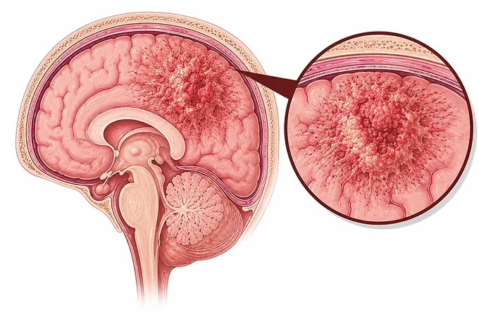
A glioma begins in the glial cells — the supporting cells that surround neurons, supply them, and keep the brain's environment stable. Because those cells are not neurons, a tumour growing from them does not simply destroy the function of the area it occupies; it infiltrates it. That single fact explains most of what makes gliomas difficult: the tumour is not a ball sitting in the brain but a region of tissue that has become abnormal, and its edge is not a border that can be seen and cut along.
-
Grade 1–4
how the tumour is expected to behave — decided by its molecular type as much as by how it looks under the microscope
-
IDH
the single most consequential marker in an adult glioma: it separates the slower-growing types from glioblastoma
-
Resection
removal as complete as can be done safely: the first treatment, and the one that shapes everything after it
-
48 hrs
from complete records to a personalized MDT plan
What a glioma is
The brain is not made only of nerve cells. Glial cells — astrocytes, oligodendrocytes and ependymal cells — outnumber neurons and do the work that keeps them functioning: supplying energy, forming the insulation that speeds up signals, clearing waste, and maintaining the fluid environment. A glioma is a tumour of those cells. Because they are distributed throughout the brain rather than confined to one place, a glioma grows within the tissue rather than pushing it aside, and that is the difference that shapes everything about how it is treated.
Three consequences follow. First, the symptoms depend on the function of the area involved: a tumour in the part of the brain that controls movement on one side causes weakness, one near the speech area causes difficulty finding words, and one in the frontal lobe may cause changes in behaviour that family notice before the patient does. Second, a seizure is often the first symptom, particularly for the slower-growing tumours, because abnormal tissue interrupts the normal electrical pattern of the cortex. Third, complete removal is rarely possible in the way it is for a tumour in an organ that can be spared, since the tumour infiltrates tissue that cannot be cut out without costing function — which is why the aim is described as the maximal safe removal rather than as a total one.
The fourth consequence is less intuitive. Because the tumour grows within the brain rather than travelling through it, gliomas almost never spread to the rest of the body. The staging that dominates other cancers — how far cells have travelled — therefore has no place here. What matters instead is the type and grade of the tumour, its molecular profile, the extent to which it could be removed, its site, and the patient's age and performance status. Those are the factors on which the plan is built, and asking for them by name is a reasonable way to understand what you are facing.
The main types
Gliomas are named for the cell they resemble and are grouped by molecular profile and age of onset. The three that account for most adult cases are defined by two tests, and it is worth knowing which one you have, because the diagnosis is not complete without it:
- Astrocytoma, IDH-mutant — arising from astrocyte-like cells and carrying a mutation in IDH. It occurs at grade 2, 3 or 4, and the grade is decided by features including the loss of CDKN2A/B. It is the type in which molecular testing most often changes what is recommended.
- Oligodendroglioma, IDH-mutant and 1p/19q-codeleted — defined by the combination of an IDH mutation with the loss of one copy each of chromosome arms 1p and 19q. It is the type that responds to chemotherapy as well as to radiotherapy, and that is the practical reason the codeletion is always tested for.
- Glioblastoma, IDH-wildtype — the commonest and most aggressive primary brain tumour in adults, and the one in which the IDH gene is not mutated. Treatment combines surgery with radiotherapy and chemotherapy, and the molecular results determine what else is appropriate.
- Circumscribed gliomas of childhood and young adults — including pilocytic astrocytoma, which grows as a defined mass rather than infiltrating, occurs mainly in children and young adults, and is often treated by surgery alone.
- Paediatric-type diffuse gliomas — a separate group with its own molecular drivers, including a mutation called H3 K27M that defines a tumour arising in the midline structures of the brain. These tumours are biologically distinct from adult gliomas and are treated differently.
- Ependymoma and other glial tumours — arising from the cells lining the fluid spaces of the brain and spinal cord, occurring at any age, and classified by site and molecular features as well as by appearance.
Grading, and what decides it
Gliomas are not staged in the way cancers elsewhere are. Instead each tumour is given a grade from 1 to 4, which describes how it is expected to behave, and a type defined by its molecular profile. The two are decided together:
| Category | In plain terms | How it is used |
|---|---|---|
| Grade 1 | A slow-growing tumour that forms a defined mass rather than infiltrating, such as a pilocytic astrocytoma | Surgery is often the whole of the treatment, with imaging follow-up afterwards |
| Grade 2 | A slower-growing tumour that has infiltrated the brain rather than stayed in one place | The molecular type decides whether treatment follows surgery straight away or whether the tumour is monitored |
| Grade 3 | A tumour that is actively growing and shows features of more aggressive behaviour | Radiotherapy and chemotherapy follow surgery, and the molecular result determines which combination is chosen |
| Grade 4 | The most aggressive group, which includes glioblastoma | Radiotherapy with chemotherapy given during it and continued afterwards, with further options considered by molecular result |
| Molecular type | Whether an IDH mutation is present, and whether 1p/19q is codeleted | These two results define which of the three adult diffuse gliomas the tumour is, and are stated as part of the diagnosis itself |
| Extent of removal | How completely the tumour was removed, assessed on the scan taken after the operation | One of the few factors within treatment's control, and a decisive influence on what is recommended next |
The point worth drawing out is that the grade is no longer decided by appearance alone. A tumour that looks relatively unremarkable under the microscope may be graded 4 because of a molecular finding, and a tumour with worrying appearance may carry a marker that places it in a better-defined group with a clearer treatment path. This is the opposite of how most patients expect a diagnosis to work, and it is the reason a report that gives only the older name of the tumour — "anaplastic astrocytoma", for example — is an incomplete report.
It also means that the diagnosis should be requested in full. Asking for the tumour type with its molecular profile, the grade and the reason for it, and the extent of removal judged on the postoperative scan is a reasonable set of questions at the outset, because each of the three changes what is offered.
The markers that guide treatment
In glioma, molecular testing is not an add-on to the diagnosis — in the adult diffuse gliomas it is the diagnosis. Four markers carry most of the decisions, and a small number of others matter in specific situations:
| Marker | What it is | How it changes management |
|---|---|---|
| IDH1 and IDH2 | A mutation in a gene involved in the cell's metabolism, found in most slower-growing adult gliomas and absent from glioblastoma | Its presence or absence defines which of the three adult diffuse gliomas the tumour is, and therefore the treatment, the follow-up and the options if it returns |
| 1p/19q codeletion | The loss of one copy each of two chromosome arms, occurring together rather than separately | Confirms oligodendroglioma, the type that responds to chemotherapy as well as to radiotherapy — which is why treatment for this group combines both |
| MGMT promoter methylation | A chemical switch that silences a gene which repairs the kind of damage the chemotherapy drug causes | Identifies the tumours most likely to benefit from temozolomide, and is reported before that decision is made |
| H3 K27M and BRAF changes | Two markers at opposite ends of the age range: H3 K27M in a tumour arising in the midline of children and young adults, and BRAF alterations in several slower-growing and circumscribed gliomas | H3 K27M identifies a biologically distinct tumour treated on its own terms; a BRAF alteration may open the possibility of a targeted drug |
Beyond those four, several results settle details. A CDKN2A/B deletion in an IDH-mutant astrocytoma is one of the findings that places it at grade 4, which changes the treatment intensity even though the tumour carries the favourable IDH mutation. ATRX loss with a TP53 mutation is characteristic of the astrocytoma group rather than the oligodendroglioma group, and helps settle the diagnosis where the codeletion result is borderline. A TERT promoter mutation and EGFR amplification are features of the glioblastoma group. In a child or young adult, the finding of a BRAF fusion or NF1 alteration may point to a targeted approach instead of conventional treatment.
The practical instruction is to ask for the molecular report itself. In this disease, more than in most, the diagnosis is a set of test results rather than a name, and a plan that states the type without the markers is a plan with its rationale removed.
Why patients come to China for glioma care
Glioma care depends on a small number of capabilities that have to work together, because the treatment sequence is long and each step depends on the last: imaging that identifies what can safely be removed, surgery performed at the edge of function without damaging it, molecular pathology reported fast enough to inform the plan, and radiotherapy delivered with precision to a target next to critical tissue.
- Molecular neuropathology available in-house — IDH, 1p/19q, ATRX, TP53, TERT, MGMT and the paediatric markers reported as part of the diagnosis, because the treatment for the three adult diffuse gliomas differs.
- Surgery planned around function — functional MRI, diffusion tractography and neuropsychological assessment before the operation, and awake craniotomy with mapping when the tumour lies near the areas controlling speech or movement.
- Techniques that increase the extent of removal — intraoperative monitoring and imaging, and fluorescence guidance, so that the boundary of the tumour can be judged during the operation rather than only afterwards.
- Precision radiotherapy — fractionated treatment shaped to the target, with attention to the dose received by the surrounding brain, and the option of re-irradiation in selected recurrences.
- Molecularly chosen drug treatment — temozolomide during and after radiotherapy, the PCV combination or temozolomide for oligodendroglioma, an oral IDH inhibitor for selected patients with a slower-growing IDH-mutant tumour, and bevacizumab for swelling in recurrent disease.
- Rehabilitation as part of the plan — physiotherapy, speech and language therapy, and cognitive support, which determine how much function is recovered after an operation rather than merely how complete it was.
Because glioma treatment is a sequence rather than an episode, the arrangements differ from those for many cancers. Patients who live elsewhere usually have the operation, the pathology review and the radiotherapy planning in China, then return home for the chemotherapy and monitoring phases under a written schedule, coming back for defined reviews or if the imaging changes. The records that make that possible are the operative and pathology reports together with the molecular panel, the postoperative MRI, the radiotherapy plan with dose and target volumes, and the drug history with dates. Send those, and the written assessment is returned within 48 hours of the file being complete.










