


Bladder Cancer Overview
Bladder Cancer Overview
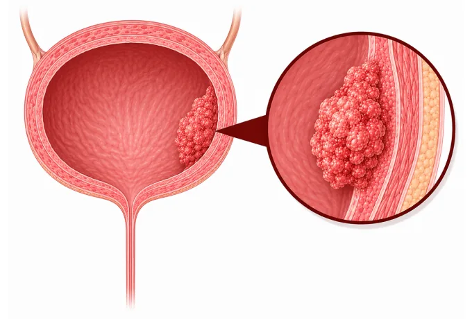
The bladder is a muscular sac lined with a specialised epithelium called the urothelium, which stretches as the bladder fills and flattens when it empties. Cancer beginning in that lining is the commonest tumour of the urinary tract, and it announces itself in the least ambiguous way any cancer can: blood in the urine that the patient can see, and that characteristically does not hurt. Painless visible haematuria is not a symptom to wait out — it is the reason cystoscopy exists.
-
~90%
of bladder cancers are urothelial carcinoma — the type that begins in the bladder's lining
-
75%
are found before the tumour has reached the muscle wall, and are treated without removing the bladder
-
Painless
visible blood in the urine is the commonest first sign — and a finding that is investigated, not watched
-
48 hrs
from complete records to a personalized MDT plan
What bladder cancer is
The bladder stores urine. Its inner surface is lined by the urothelium, a layer of transitional cells that is unusual in two ways that shape this disease. It is in direct contact with whatever the urine contains, so it is exposed for decades to the carcinogens the body is excreting — which is why smoking and industrial chemicals cause this cancer more than most. And it runs continuously from the kidney, down the ureter, through the bladder and out along the urethra, so a field of urothelium can develop tumours in more than one place.
That second feature explains the shape of the diagnostic pathway. Because the lining is a single continuous surface rather than an isolated organ, finding a bladder tumour prompts imaging of the upper urinary tract as well — and follow-up cystoscopy is scheduled for years, because a new tumour can appear in the same lining at a different site.
The decisive question at diagnosis is not how large the tumour is but how deep it has grown. The bladder wall has a lining, a layer of connective tissue beneath it, and then the thick muscular layer that contracts to empty the bladder. A tumour that has not reached the muscle — non-muscle-invasive disease, which is about three-quarters of cases at diagnosis — is removed through the urethra and then treated by putting drugs directly into the bladder. A tumour that has reached the muscle has a different natural history and is treated either by removing the bladder or by irradiating it together with chemotherapy. Almost everything in this cluster follows from that one division.
The main types
The type of bladder cancer is defined by the cells it arises from, and the type determines both its behaviour and whether the usual treatments apply:
- Urothelial carcinoma — about nine in ten cases, arising in the transitional lining and growing either as a papillary frond or as a flat lesion. The flat, high-grade form — carcinoma in situ — is the one that causes irritative symptoms rather than bleeding, and it is the form most likely to progress.
- Squamous cell carcinoma — arises from the lining after years of chronic irritation or inflammation, including long-standing catheters, repeated infection and infection with Schistosoma haematobium. It is more common where schistosomiasis is endemic and tends to be diagnosed at a later stage.
- Adenocarcinoma — rare, gland-forming, and sometimes arising from the remnant of the urachus, the embryonic channel that once connected the bladder to the umbilicus.
- Small-cell and neuroendocrine carcinoma — uncommon and aggressive, treated with chemotherapy-based protocols rather than by surgery alone.
- Histological variants of urothelial carcinoma — micropapillary, nested, plasmacytoid and sarcomatoid patterns, reported alongside the main diagnosis because they behave more aggressively and are more often managed with early cystectomy.
Because urothelial carcinoma dominates, most patients are dealing with it. The other types matter for two practical reasons: squamous and adenocarcinoma may not respond to the intravesical drugs used for urothelial disease, and the aggressive urothelial variants are a reason to move to cystectomy sooner rather than to extend bladder-preserving treatment.
What each stage means
Staging combines the AJCC classification with the risk group that clinicians use to choose treatment. For bladder cancer the risk grouping is doing most of the work, because two tumours with the same stage number can be managed quite differently depending on grade and history:
| Category | In plain terms | How it is usually approached |
|---|---|---|
| Non-muscle-invasive, low risk | A small, solitary, low-grade papillary tumour on the surface lining | Complete resection through the urethra, plus a single immediate instillation of chemotherapy into the bladder |
| Non-muscle-invasive, intermediate risk | Larger, multiple or recurrent tumours that have not reached the muscle | Resection followed by BCG immunotherapy, given as induction and then maintenance |
| Non-muscle-invasive, high risk | Tumours that have reached the connective tissue layer, high-grade tumours, or carcinoma in situ | BCG, with radical cystectomy offered early and discussed as the definitive option |
| Muscle-invasive, localised | The tumour has reached the muscle wall, with no spread beyond the bladder | Chemotherapy before surgery, then removal of the bladder with lymph node dissection; or chemoradiotherapy in a bladder-preserving plan |
| Node-positive | Cancer found in the pelvic lymph nodes | Systemic treatment with chemotherapy or an antibody-drug conjugate combination, with surgery or radiotherapy in selected cases |
| Metastatic | Spread to distant nodes, bone, lung or liver | An antibody-drug conjugate with immunotherapy, or platinum-based chemotherapy with maintenance treatment |
Two features of that table are worth drawing out. The three non-muscle-invasive rows are one stage but three different diseases: a low-risk papillary tumour may need no more than a resection and one dose of chemotherapy, while a high-risk tumour that has reached the connective layer carries a real chance of progressing to muscle-invasive disease and is a reason to discuss cystectomy early, before it does. And the last three rows overlap: a tumour that has reached the muscle can still be cured in the bladder by the combined approach, whereas once disease is metastatic the aim shifts to controlling it systemically.
Because the risk category depends on the resection specimen, the pathology report is the document that decides the plan — and specifically whether the specimen contains muscle. A resection that does not sample muscle cannot tell whether the tumour is muscle-invasive, which is why an incomplete first resection is often repeated rather than treated as a final answer.
The markers that guide treatment
Bladder cancer is not usually described by its mutations in ordinary conversation, but a short list of markers now determines who can receive which treatment, and testing is done at the point where it changes a decision:
| Marker | What it is | How it changes management |
|---|---|---|
| FGFR3 alteration | Activating changes in the FGFR3 gene, found in a substantial share of lower-grade tumours | Marks a biology that is less likely to progress; in advanced disease an FGFR inhibitor becomes a targeted option instead of chemotherapy |
| Nectin-4 | A protein on the surface of most urothelial cancer cells | The target of enfortumab vedotin, an antibody-drug conjugate used from first line in advanced disease |
| PD-L1 and mismatch repair status | Markers of how the tumour interacts with the immune system | Guides immune checkpoint inhibitor treatment and identifies tumours that can respond to immunotherapy |
| DNA repair genes | Faults in genes such as ERCC2 and BRCA1 or BRCA2 that repair the damage chemotherapy causes | Studied as a way of judging how well cisplatin-based chemotherapy is likely to work, and explored for guiding platinum versus non-platinum choices |
The practical point is about timing. For non-muscle-invasive disease, none of these tests changes the immediate plan, which is resection followed by intravesical treatment — so they are not routinely ordered. For advanced disease the position is the opposite: Nectin-4 and PD-L1 testing decide whether the current first-line combination is appropriate, an FGFR3 result decides whether a targeted drug is available instead, and testing for DNA repair faults is relevant when platinum chemotherapy is being considered. Ask which of these has been done before the systemic plan is fixed.
Why patients come to China for bladder cancer care
Bladder cancer care is decided by four capabilities, and a service either has them or does not: reliable cystoscopy and complete resection, intravesical treatment given on an unbroken schedule, surgery and radiotherapy that can offer both cystectomy and bladder preservation, and systemic options that are current.
- Blue-light cystoscopy and complete resection — imaging the bladder with a photosensitising agent reveals flat lesions that ordinary white-light cystoscopy can miss, which is exactly the kind of lesion most likely to be high grade.
- Intravesical therapy delivered on schedule — BCG induction and maintenance is a regimen rather than a single treatment, and its value depends on the maintenance doses actually being given on time.
- Radical cystectomy with urinary reconstruction — removal of the bladder with lymph node dissection, and a choice of diversion: an ileal conduit or an orthotopic neobladder constructed so that the patient can continue to urinate normally.
- Bladder-preserving chemoradiotherapy — radiotherapy given with a radiosensitising drug after a complete resection, for patients who cannot undergo cystectomy or who elect to keep the bladder, with cystoscopy follow-up built into the plan.
- Current systemic treatment — antibody-drug conjugate combinations, platinum-based regimens and immune checkpoint inhibitors for advanced disease, with marker testing arranged in-house.
- Structured surveillance — cystoscopy on a fixed schedule for years, because a new tumour in the same urothelium is a normal part of this disease rather than a failure of the first treatment.
Two practical consequences follow. Because the choice between cystectomy and bladder preservation depends on tumour features, on fitness for surgery and on the patient's own priorities, a written assessment should set out both routes rather than one — and should say which of them the pathology supports. And because surveillance is lifelong, the follow-up schedule belongs in the plan from the beginning. Send the resection pathology report, the imaging and the treatment history, and the written assessment is returned within 48 hours of the file being complete.










