


Arteriovenous Malformation (AVM) Overview
Arteriovenous Malformation (AVM) Overview
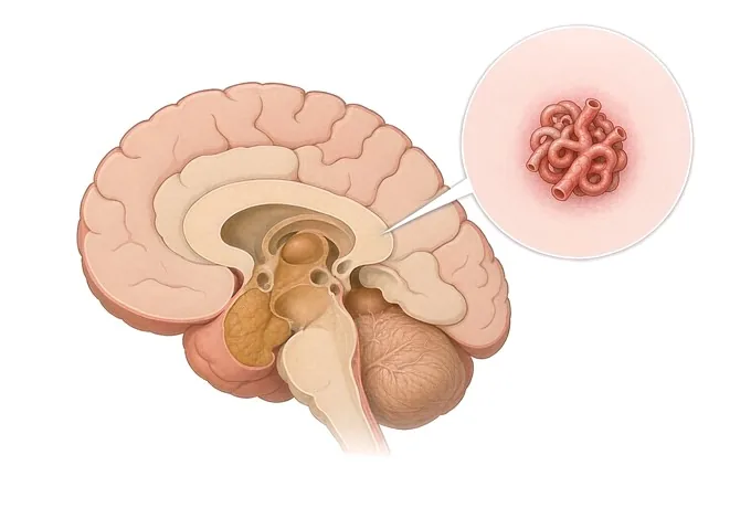
A brain arteriovenous malformation is a developmental abnormality of the blood vessels: an artery connects directly to a vein through a tangle of vessels, without the capillary bed that normally sits between the two. It is usually present from birth, and it is uncommon — most published estimates place it well below one per cent of the population. Because the capillary bed is missing, blood crosses at arterial pressure into veins that were not built to carry it. That is the whole of the problem, and it explains everything the malformation can do: it can bleed, it can irritate the surrounding brain and cause seizures, and it can divert blood away from tissue that needs it.
-
National
Stroke Center construction unit, and a national GCP base
-
3D DSA
Alongside 3.0T MRI, dual-source CT, PET-CT and 24-hour video EEG
-
48 hours
Written second-opinion report after complete records arrive
-
124+
Countries and regions our international patients come from
What an AVM is, and the shortcut in the circuit
A normal capillary bed is a pressure-reducing device. Blood leaves an artery at arterial pressure and arrives at a vein at venous pressure because it has been passed through a very large number of very small vessels along the way. In an arteriovenous malformation that section of the circuit is missing. An artery feeds directly into a tangle of abnormal vessels — the nidus — which drains straight into a vein.
Two consequences follow, and they account for every symptom the malformation produces. The first is pressure: the veins on the far side of the nidus are carrying blood at a pressure they were not built for, and over years they dilate and their walls remodel. The second is flow: the shunt is a low-resistance path, and the circulation takes it. Blood that would have supplied the brain around the nidus is drawn through the shortcut instead.
The malformation is described in three parts, and a report worth having names all three: the feeding arteries that supply it, the nidus itself, and the draining veins that carry blood away. Which arteries feed it and how the vein drains are not anatomical trivia — they are two of the three items that make up the grading scale, and they are what determines whether the malformation can be removed.
One thing should be said early, because it changes how the rest is read. An AVM is usually present from birth and not inherited in the ordinary sense: it arises during the development of the vessels rather than being passed down. The exceptions are specific and uncommon, and they are dealt with in the causes page. For the great majority of patients, nothing they did caused it, and nothing they did would have prevented it.
Size, drainage and eloquence — the grade
An AVM is not judged by its size alone. Three features are scored separately and added together, and the total is what surgeons actually discuss.
Size is measured across the largest dimension of the nidus, on angiography. Under 3 cm scores one point, from 3 to 6 cm scores two, and above 6 cm scores three. The categories are wide because size is a proxy for something more practical: how much brain has to be traversed to reach the malformation, and how much blood is moving through it.
Eloquence describes where it sits. A malformation adjacent to brain that serves language, movement, vision or sensation is called eloquent, and scores a point; one in a region where a deficit would be silent does not. This is the component patients find hardest, because it is the one where two malformations of identical size are entirely different problems.
Venous drainage is the third. A malformation that drains only through veins on the surface scores nothing; one that also drains through the deep venous system — the internal cerebral veins, the basal vein, the vein of Galen and their tributaries — scores a point. Deep drainage is not merely a marker of depth: it usually means that the malformation is supplied by perforating arteries that are difficult to control, and it is the component most associated with a worse outcome from surgery.
Size, drainage and whether it has bled
Three axes carry most of the decision. One is anatomical, one is the pattern of drainage, and one is simply whether blood has already escaped — because that last one changes what the risk of waiting actually is.
| Axis | How it is described |
|---|---|
| Whether it has bled | Unruptured, or previously ruptured. This is the axis that most changes the arithmetic: bleeding once is the factor most consistently associated with bleeding again, and the randomised evidence that favours conservative management enrolled only unruptured malformations. A malformation that has already bled is a different proposition from one that has not, even at the same size and in the same place. |
| Size of the nidus | Scored as under 3 cm, 3–6 cm, or above 6 cm on the largest dimension measured at angiography. Size matters for what it implies about access and about flow, and it is one of the three components of the surgical grade. It is not a predictor of bleeding on its own: small malformations bleed, and the association between size and rupture risk is weaker than most patients expect. |
| Eloquence of the surrounding brain | Whether the malformation lies next to tissue serving language, movement, vision or sensation. Two malformations of the same size can be graded a point apart on this alone, and the difference is between a lesion that can be removed with an acceptable deficit and one where removal would cost a function the patient cannot spare. |
| Pattern of venous drainage | Superficial only, or with a deep component. Deep drainage usually indicates supply from perforating arteries that are difficult to control, and it is the component most consistently associated with a worse surgical outcome. It is also the feature that pushes a small, deep, eloquent malformation towards radiosurgery rather than towards an operation. |
| Associated aneurysms | An aneurysm on a feeding artery, within the nidus, or on an unrelated vessel. When a malformation has bled, this matters directly: the aneurysm may be the source rather than the nidus, and the two are not treated in the same way. Reports should state whether one has been looked for and what was found. |
| How it presented | Found after a bleed, after a seizure, during investigation of a headache, or by chance on a scan taken for something else. Presentation determines the urgency and, in the first instance, which investigation comes first. Seizures and headaches are not warnings of an imminent bleed; they are reasons the malformation was found. |
| Stage of treatment, and time since it | Untreated, partially embolised, or treated and awaiting confirmation. Radiosurgery has a latency of one to three years during which the malformation is still present and can still bleed, so the time since treatment is part of the risk rather than a footnote. Obliteration is confirmed on an angiogram, not inferred from the absence of symptoms. |
Read together, these axes do not answer "how dangerous is this AVM". They answer which of three situations this is: a low-grade malformation where removal is curative and the risk of surgery is lower than the lifetime risk of leaving it; a malformation where the risk of treating it exceeds the risk of watching it, and watching is the treatment; or one where no single technique is enough and the plan is a sequence staged over months.
How an AVM is mapped
The order depends on how the malformation declared itself. After a suspected bleed the sequence is urgent and starts with CT; otherwise the question is which study gives the anatomy in enough detail to assign a grade.
| Investigation | What it settles |
|---|---|
| CT of the head, then CT angiography | Whether blood is present, where it lies, and how much. The same scan shows the malformation's vessels in outline and, importantly, whether the bleeding is into the brain tissue, into the ventricles, or into the space around the brain — which points towards or away from an associated aneurysm as the source. |
| MRI and MR angiography | Where the nidus sits in relation to the brain around it, and whether there is old blood — evidence of a bleed the patient never knew about. It is the study most often used to follow an untreated malformation, because it involves no ionising radiation. |
| Digital subtraction angiography | The reference study, and the one the plan is built on. It shows the feeding arteries filling, the nidus, and then the veins, in sequence and in time — which is the only way to tell arterial from venous phases. It is what the three-component grade is assigned on, and it is performed by the team that would carry out the treatment. |
| Functional mapping | Where language, movement and sensation sit relative to the malformation, by functional MRI or by another mapping technique. Used when the lesion is near eloquent cortex, and it can change a plan from one that assumes a deficit to one that avoids it. |
| Catheter angiography after treatment | Whether the malformation has been obliterated. This is the only reliable way to say so: after radiosurgery in particular, the vessels can look closed on MRI while a channel remains. Until an angiogram shows obliteration, the risk of bleeding has not been removed. |
A report worth having therefore answers six things: how big the nidus is, which arteries feed it, how it drains, whether it sits in eloquent brain, whether there is an associated aneurysm, and whether it has bled before — the last of which is answered as often by the MRI as by the history.
Why patients travel to China for AVM treatment
A patient whose malformation has just bled needs the nearest hospital able to treat it, and no one should travel during one. Travel becomes reasonable afterwards, and in three situations: when an unruptured malformation has been found and the question of whether to treat it at all needs a second opinion; when a malformation has been treated and the follow-up question is whether obliteration has been achieved; and when the work ahead is rehabilitation after a bleed.
Our neurology and neurosurgery services are organised as a standardised stroke centre built to national stroke-centre standards, and are a National Stroke Center construction unit and national GCP base. Cerebrovascular cases are planned jointly by neurology, neurosurgery and rehabilitation rather than handed from one department to the next, and interventional work — the treatment of arteriovenous malformations, aneurysms, Moyamoya disease and arterial stenosis — is carried out on 3D DSA, alongside 3.0T MRI, dual-source CT, PET-CT and 24-hour video EEG.
The practical questions are answered in the same way as for any other condition we treat. Records reviewed by a neurovascular specialist receive a written opinion within about 48 hours of complete records arriving, and the opinion states what the images show, what grade the malformation falls into, which of the three techniques is reasonable here and which is not, and what remains uncertain. Complete treatment in China typically runs at 30–50% of comparable care in Europe or the United States. We work with more than 30 commercial insurers for direct billing and have received patients from 124 countries and regions. Where the question is whether an unruptured malformation should be treated at all, that is usually answerable from the images, and a video consultation is a reasonable first step rather than a journey.










