


Adenomyosis Overview
Adenomyosis Overview
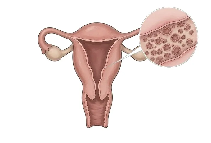
Adenomyosis is the presence of tissue that resembles the lining of the womb within the muscle of the womb itself. It is not cancer and it is not an infection. What makes it matter is that this tissue behaves as the lining behaves: it thickens and breaks down with the hormonal cycle, but it is embedded in muscle, so the blood it produces has nowhere to go. The muscle around it responds by becoming thicker and less efficient, and the womb becomes enlarged, bulky and tender. That single fact — the tissue is inside the wall rather than on a surface or free in the pelvis — explains almost everything about the condition. Periods become heavier. Pain that used to be limited to the period becomes worse year by year rather than staying the same. The womb itself can be felt to be enlarged on examination. Two distinctions run through everything that follows. Adenomyosis and endometriosis are different conditions that are constantly confused, because they share the cyclical pain and the heavy bleeding while sitting in different places and answering to different treatments. And how much disease is seen on a scan is a poor guide both to how much it is affecting someone and to what should be done about it.
-
The bleeding tends to get heavier, and the pain worse
an increase over years rather than a stable pattern is what separates this from ordinary period pain
-
Adenomyosis and endometriosis are two different conditions
one sits inside the wall of the womb and one outside it; the symptoms overlap, the treatments do not
-
The amount of disease on a scan is a poor guide to the plan
what changes the plan is the bleeding, the pain and whether a pregnancy is wanted
-
Removal of the womb is one complete option among several
it is the only treatment that removes the disease with the womb, and it is not the only treatment that works
What adenomyosis is, and what the name gets wrong
It is a condition defined by where the tissue is, and the position of the tissue explains the symptoms:
- The tissue is inside the muscle of the womb. It is not in the cavity and it is not outside the womb in the pelvis. It is embedded in the wall that is supposed to contract and squeeze shut during a period, and that is why the bleeding and the pain become the two problems the condition is known by.
- The womb becomes enlarged, bulky and tender. The muscle around the embedded tissue thickens, and the whole organ grows. It is often felt as a boggy, tender uterus on examination, which is a real finding and not a description of how the patient feels.
- The periods become heavier, and the pain becomes worse over time. This direction of travel is the feature that separates the condition from ordinary period pain, which usually appears soon after the periods begin and then stays roughly the same.
- It is not the same condition as endometriosis. Endometriosis is tissue like the lining of the womb growing outside the womb — on the peritoneum, an ovary, the bowel or the bladder. Adenomyosis is the same kind of tissue growing inside the wall of the womb. Both cause cyclical pain and heavy bleeding, both are coded separately, and a woman can have both, but the disease being treated sits in a different place and answers to different measures.
- Many women with it have no symptoms at all. It is regularly found when imaging is performed for an unrelated reason, or when the womb is examined after removal for something else. Finding it by accident is not the same as being told it is unimportant: it changes what is watched, and it changes what a later symptom will be attributed to.
What the name does not mean. The word describes two things and both are worth knowing: adeno refers to glandular tissue like the lining of the womb, and myosis refers to muscle. It does not mean cancer, and it does not turn into cancer. It does not mean the womb has to be removed. It does not mean the pain is imagined or that it is simply what periods are like at this age. It does not mean fertility is over. What it means is that tissue resembling the lining of the womb is present where it does not belong, that its position is the reason for the bleeding and the pain, and that there is a sequence of treatments to work through rather than a single answer.
The five forms it takes, and what each one changes
Where in the wall the tissue sits, and how much of the womb is involved, determines what the symptoms are like and which treatment is worth trying first:
| What is found | What follows from it |
|---|---|
| Diffuse adenomyosis, spread through the muscle | The commonest pattern. The womb is uniformly enlarged and boggy, and no single area can be removed. Treatment works on the symptoms and on the whole womb rather than on one deposit |
| Focal adenomyosis, also called an adenomyoma | A localised area within the muscle. It can look very like a fibroid on a scan, which is one reason the two are confused, and it is the form most likely to be suitable for a womb-preserving operation |
| Disease of the junctional zone | The inner layer of muscle next to the cavity. It is the form most closely tied to heavy bleeding and to pain that starts before the period, and thickening of this layer is one of the findings imaging is read for |
| Adenomyosis with fibroids as well | Common, and it matters because the two are told apart before treatment rather than assumed. Fibroids distort the cavity and can be shelled out; adenomyosis cannot, so what each is contributing changes which operation is offered |
| Adenomyosis with endometriosis as well | The cyclical pain is shared, and the two are planned separately. Treatment aimed at the disease outside the womb will not reach the disease in its wall, so a plan that treats only one of them is a plan that leaves half the pain |
Why the form matters more than the size. A womb that is only slightly enlarged can cause bleeding that dominates a life, and a markedly enlarged womb can be found in someone whose periods are unremarkable. Focal disease can be removed while the womb is kept, and diffuse disease cannot be removed at all without removing the womb. Which of these is present is therefore the first thing established, because it decides which of the options are even available.
What changes the plan, and how the pace is set
What changes the plan is not how large the womb measures but what the condition is doing and what is wanted next:
| What changes the plan | What it changes |
|---|---|
| How much the bleeding is affecting daily life | It decides whether the first step is a measure that reduces bleeding, and it is the measure the plan is reviewed against. Bleeding that is tolerated is not the same as bleeding that is harmless, and the blood count is checked rather than assumed |
| Whether the pain is confined to the period or present most days | Pain that has stopped following the cycle points at a different balance between active disease and the changes it has caused, and it shifts what is likely to help |
| Whether a pregnancy is wanted now, later, or not at all | It decides which options are on the table at all. Methods that suppress the cycle control the symptoms and prevent pregnancy; a womb-preserving operation is planned around preserving fertility; removal of the womb ends the question |
| The size and the shape of the womb | It decides whether a womb-preserving operation is technically possible and whether an intrauterine system can be fitted and is likely to stay in place |
| Whether fibroids or endometriosis are present as well | It decides whether more than one problem is being treated, and whether the operation needs to be planned with the disciplines that would be needed for both |
Why pace is the practical question. Patients who ask how serious the condition is usually mean how quickly they should move and what to agree to first. Those answers come from how much the bleeding is costing in daily life, how much of the month the pain takes, whether a pregnancy is on the horizon, and how long a treatment needs before it can be judged. That is why the plan is written as a sequence with review dates rather than as a single decision.
What the assessment is looking for
The assessment takes the bleeding history, takes the pain history, examines the womb and uses imaging for what it can show:
| What is assessed | What it establishes |
|---|---|
| The bleeding history, taken as a change over time | How the periods differ from a few years ago, how many days of heavy flow there are, whether clots are passed and whether bleeding occurs between periods, which together carry much of the diagnosis |
| The pain history | Whether the pain has worsened year by year rather than stayed level, whether it outlasts the bleeding, and whether it is present on days without bleeding, which is the pattern that separates it from ordinary period pain |
| A pelvic examination | Whether the womb is enlarged, bulky and tender, whether it is fixed, and whether there is any other explanation for the findings, all of which change what is suspected |
| A transvaginal ultrasound | The changes of adenomyosis within the muscle of the womb, and at the same time the size of the womb, the presence of fibroids and of polyps, and any ovarian findings |
| An MRI where it will change something | The distribution of the disease, the thickness of the inner muscle layer and the relationship to fibroids, used to decide whether a womb-preserving operation is possible and how it should be planned |
| A blood count and the alternatives | Whether years of heavy bleeding have produced anaemia that needs treating, and whether another cause such as a polyp, thyroid disease or a bleeding disorder is present, because each is treated differently |
Why the assessment stops when it has answered the question. Not every woman needs an MRI, and nobody needs repeated imaging to watch a condition that imaging does not track. The assessment is finished when the bleeding has been measured, the pain has been characterised, the womb has been examined, and the other explanations that would change the treatment have been excluded.
Why patients come to China for this assessment
This is a condition whose treatments differ in what they cost a woman, and travelling for it is usually about having the options set out together:
- The bleeding is measured rather than assumed. How much is being lost, whether it has produced anaemia, and what would count as an improvement are written down, so that the effect of a treatment can be judged rather than guessed at.
- The womb is examined and imaged properly. The examination is performed as part of the assessment, and ultrasound is read specifically for the changes of adenomyosis and for the fibroids that imitate it, rather than being reported as normal because nothing else was sought.
- The options are set out in sequence, including the ones that preserve the womb. Reducing the bleeding, suppressing the cycle, a womb-preserving operation, embolisation and removal of the womb are all discussed before a plan is written, rather than one of them being presented as the only answer.
- Fertility is treated as a factor rather than an afterthought. Whether a pregnancy is wanted now, later or not at all is established before the plan is made, because it decides which of the options is available at that moment.
- The plan and the records are prepared to travel. Results are issued with their dates and the technique used, and the written plan names what is being treated, what is being watched, when the next review is, and what would change the approach.
What this does not promise. A completed assessment does not guarantee that the bleeding will stop or that the pain will go, and it does not promise that a pregnancy will follow. What it does is establish how much is being lost, what is causing it, which options preserve the womb and which do not, and what the order of trying them should be. Saying which parts can be acted on and which cannot is part of the assessment rather than a caveat added afterwards.










