2026-07-30
When an accident robs an ordinary person of their sight, an acquired blind person is born. From then on, the eyes seem to be covered with several layers of gauze, able only to perceive a blurry patch of light. "Mom, why can't I see?" Until one day, even that patch of light disappears. "Mom, where are you?" For them, blindness is like a movie that suddenly goes black halfway through, with only the lingering negatives of the past remaining in their minds. They can only recall the colorful scenes in the long darkness that follows
The eyes are the windows to the soul. If vision is damaged, the blow to a person is immense—even devastating.
Aggressive pituitary adenomas are exactly the kind of brain tumor that can cause acquired blindness. What is an aggressive pituitary adenoma? How is it treated? Are there relevant case studies? Bookmark this article to fully understand aggressive pituitary adenomas.
What is an aggressive pituitary adenoma?
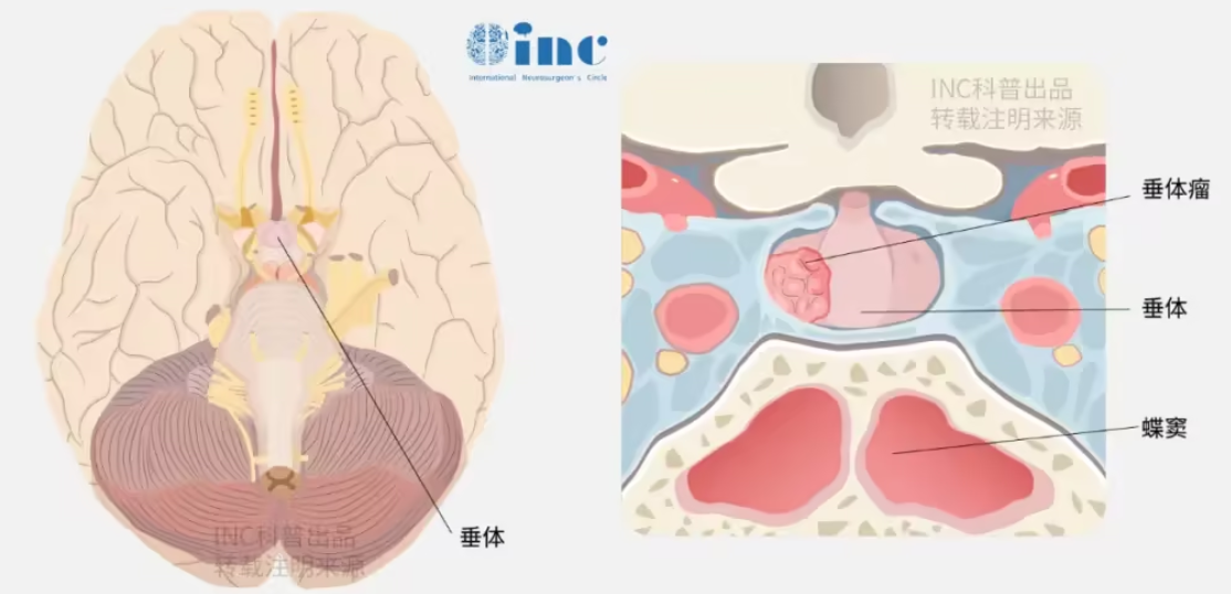
Pituitary adenoma (PA) is one of the most common benign intracranial tumors, accounting for about 15% of intracranial tumors.
Clinical studies have found that some pituitary adenomas with Knosp grade 3 or 4 exhibit aggressive characteristics. Tumor growth often invades adjacent tissues, such as the dura mater, skull base bone, encases the internal carotid artery, and compresses the optic nerve. Aggressive pituitary adenomas have a low total resection rate, poor response to radiotherapy and drug therapy, and a high recurrence rate, making them one of the more difficult neurosurgical diseases to treat clinically.
Aggressive pituitary adenoma – Knosp grading
Table 1: Modified Knosp grading for pituitary adenomas
| Grade | Description |
|---|---|
| 0 | Tumor confined to the sellar region, no extension into the cavernous sinus |
| 1 | Tumor extends into the medial cavernous sinus, but does not cross the medial tangent line of the internal carotid artery |
| 2 | Tumor extends into the superior or inferior cavernous sinus, but does not cross the lateral tangent line of the internal carotid artery |
| 3 | Tumor extends lateral to the internal carotid artery, occupying the lateral compartment of the cavernous sinus |
| 4 | Tumor completely encases the internal carotid artery |
Table 2: Imaging markers of aggressive pituitary adenomas
| Marker | Finding |
|---|---|
| Irregular shape | Invasion of adjacent structures |
| Bone erosion | Skull base bone destruction |
| Rapid growth | Significant growth on serial imaging |
| Invasion of cavernous sinus | Encasement of carotid artery |
Can aggressive pituitary adenomas be treated with endoscopy?
In addition to traditional microscopic craniotomy, with the development of neurosurgery and continuous updates in medical equipment, neurosurgical procedures are gradually moving toward minimally invasive approaches. Most pituitary adenomas can be resected via an endoscopic endonasal transsphenoidal approach. This greatly reduces surgical trauma, lowers blood loss, allows faster postoperative recovery, and achieves better surgical outcomes, making total tumor resection possible.
Advantages of neuroendoscopy for vision protection
Endoscopic endonasal surgery via the ethmoid approach lateral to the middle turbinate preserves the nasal septum, middle turbinate, and sphenoid sinus, with almost no impact on nasal physiological function. Patients can be discharged three days after surgery. Neuroendoscopy is not limited by depth or narrowness; it allows close‑up observation with a clear field of view. Combined with neuronavigation, it offers significant advantages.
The optic nerve canal is located posterior to the ethmoid labyrinth and adjacent to the internal carotid artery. If the labyrinth is injured during endoscopy, it can be life‑threatening. Therefore, combining neuroendoscopy with neuronavigation compensates for each other's shortcomings.
However, the nasal cavity is a long, narrow passage, and instruments can interfere with each other, affecting technique. After entering the sphenoid sinus, the freedom of bimanual operation improves significantly.
Many experts require a designated assistant to hold the endoscope to obtain good visual exposure and ensure smooth surgery. The assistant must fully understand the lead surgeon's intentions, maintain good hand stability, and be willing to make some sacrifices to hold the endoscope. It often takes a long time for the assistant and lead surgeon to coordinate and achieve a good working relationship.
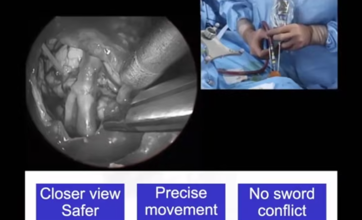
Professor Sébastien Froelich, an international skull base surgery expert and master of neuroendoscopy, invented the "chopsticks technique" for neuroendoscopy—a good solution!
During the nasal phase, mucosal flap creation, sphenoid sinus opening, and sellar floor exposure, the chopsticks technique allows effective bimanual operation with three instruments. After entering the subdural space, we usually switch to a two‑person, three‑hand operation for better stability.
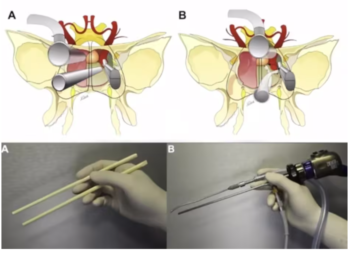
As proficiency with the chopsticks technique improves, the opportunities to enter the subdural space increase, allowing two‑person, four‑hand or even five‑hand operations, taking full advantage of endoscopic tumor resection. Sometimes it plays an unexpected role in specific situations. At the same time, training young doctors in the chopsticks technique enables faster skill acquisition.
Case study of an aggressive pituitary adenoma – A 16‑year‑old girl with recurrence 8 years after surgery, facing blindness: can re‑operation restore vision?
A 16‑year‑old girl had a large aggressive pituitary adenoma compressing the ophthalmic artery, optic nerve, and other critical structures, causing vision loss that was unresponsive to medication. Why was this "benign" tumor so severe? After endoscopic resection, her vision improved. However, 8 years later, the disease returned. The tumor recurred and her vision gradually deteriorated. Could a second surgery be performed safely and restore her vision?
A 16‑year‑old girl with vision loss – the diagnosis was a large pituitary adenoma!
At age 16, Xiaokui could no longer see clearly—textbooks, classmates' faces... it seemed she had become "nearsighted." Her right eye vision worsened to only 0.1. What does that mean? A visual acuity of 0.1 indicates very poor sight; she could only see the largest line on the eye chart. Moreover, she suffered from persistent headaches and irregular menstruation.
After consulting a neurosurgeon, an MRI revealed a pituitary prolactinoma (Figure 1). The reason for her severe vision loss was tumor compression of the optic nerve. As the tumor continued to grow, she would likely lose her vision permanently…
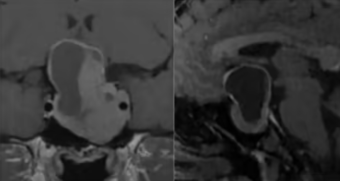
Figure 1: Initial lesion
Failed medical therapy, first surgery provided temporary relief
Clinically, prolactinomas respond well to medical therapy alone as first‑line treatment compared to other pituitary tumors. Initially, Xiaokui chose medication, taking cabergoline daily. However, under cabergoline treatment, the tumor volume did not change significantly, and medical therapy failed.
Subsequently, Xiaokui underwent surgery – an endoscopic endonasal resection. The tumor was partially removed, relieving brain compression and partially restoring vision. However, because the tumor was not totally resected, doctors recommended Gamma Knife radiosurgery. The Gamma Knife specialists determined that the residual tumor still compressed the optic nerve and was unsuitable for Gamma Knife, leaving her at risk of recurrence at any time.
Eight years later, recurrence was inevitable – sudden stroke and secondary empty sella syndrome
Eight years later, Xiaokui developed progressive vision loss, ptosis (drooping eyelid), and was hospitalized for a sudden cerebral hemorrhage. Imaging revealed tumor recurrence, and she underwent another surgery. However, residual tumor remained. Four months later, she was referred to neurosurgery due to worsening vision. MRI showed residual tumor and secondary empty sella syndrome with downward displacement of the anterior communicating artery complex (Figure 2). Secondary empty sella syndrome occurs when a defect in the diaphragma sellae or pituitary atrophy allows the subarachnoid space to herniate into the sella under cerebrospinal fluid pressure, enlarging the sella and compressing the pituitary, leading to clinical manifestations such as sellar enlargement, visual impairment, and visual field defects.
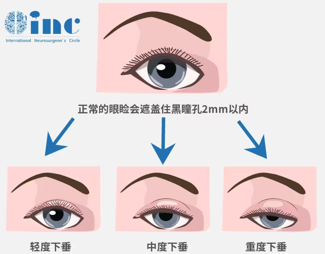
Ptosis
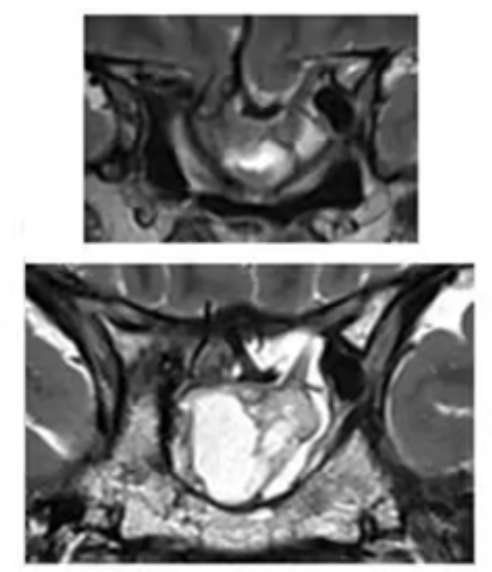
Imaging: initial tumor resection 8 years prior, recurrence with cerebral hemorrhage and sellar floor depression
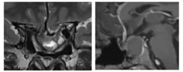
Figure 2: Preoperative MRI showing residual tumor after the second surgery and imaging features of secondary empty sella syndrome
Secondary empty sella syndrome is a problem…
The doctors explained that, in addition to residual tumor, secondary empty sella syndrome is a relatively complex condition. The resulting hypopituitarism and vision loss are chronic and progressive. During this process, the pituitary gland and optic chiasm become compressed, thinned, and displaced posteriorly and inferiorly; the small feeding vessels are also stretched and narrowed. Surgical techniques for empty sella syndrome are rarely reported. In the existing literature, the complication rate after surgery for empty sella syndrome is relatively high. Because the optic nerve pathways and chiasm have been stretched by the tumor for a long time and are adherent, postoperative scarring may cause downward traction of the opened diaphragma and chiasm, gradually increasing the degree of traction, which explains the delayed vision loss often seen months to years after surgery.

Surgical options include craniotomy, endonasal surgery, extradural reduction, and subdural reduction. If reduction is too rapid or excessive, rapid displacement of the chiasm may occur, with tearing of small vessels, leading to irreversible severe complications such as intracranial hemorrhage and rapid vision loss (as reported in the literature).
Xiaokui was well aware of the dangers of her disease, and her condition was so complex that she wanted to give herself every possible chance. Through literature and books, she learned that in this field, a French skull base surgery professor has unique advantages—Professor Sébastien Froelich, current Chairman of the WFNS Skull Base Surgery Committee and a member of the INC World Advisory Neurosurgical Group. He is a leading pioneer in international neuroendoscopic surgery, and he invented the highly efficient "chopsticks technique."
A "gleam of hope" in the darkness
"The first step is an endoscopic endonasal approach for chiasmapexy (fixation of the optic chiasm). The second step is to remove the tumor and reconstruct the sellar floor. This will prevent recurrence while alleviating the current vision loss." At Lariboisière Hospital, Professor Froelich patiently explained his treatment plan.
Surgical procedure: Because of significant adhesion and kinking between the cisternal segment and the intracanalicular segment of the optic nerve, an endoscopic endonasal chiasmapexy was performed for fixation and protection (Figure 3). The proximal optic canal was drilled to remove bone, and then the residual tumor was resected. The craniotomy was closed with artificial dura mater, watertight sutures were applied to prevent CSF leakage. The optic nerve was visualized through the diaphragm in the optic chiasmatic cistern, and abdominal fat was used to pack the sella. The sellar floor was reconstructed using nasal septal bone and titanium mesh.
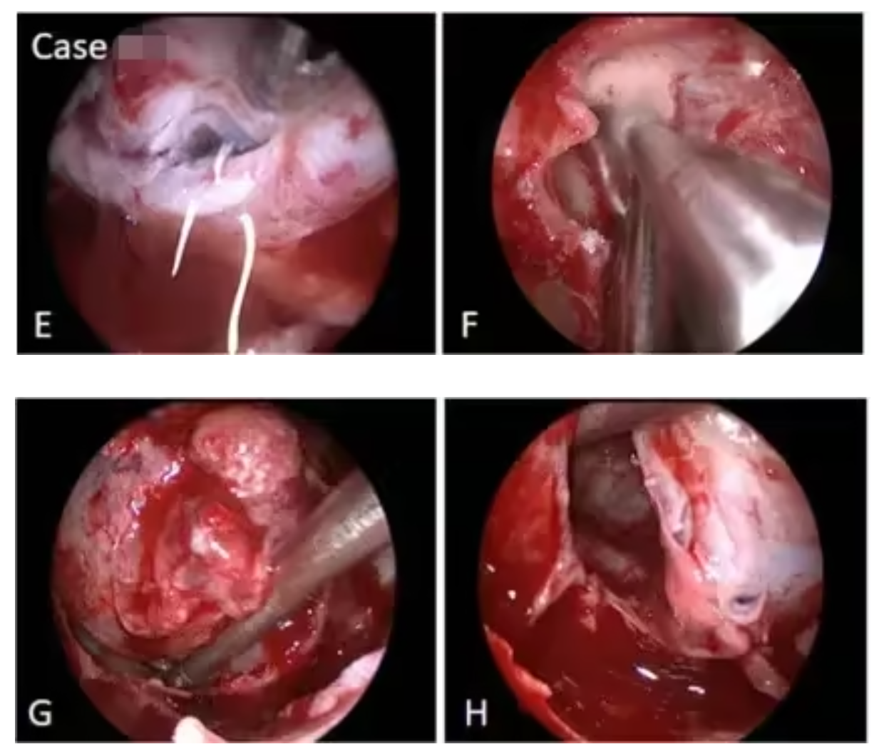
Figures 3 (e,f,g,h): (e) Intrasellar herniation of the anterior communicating artery complex; (f) drilling of the right optic canal; (g) extradural dissection of the intrasellar tumor; (h) intradural tumor resection.
One year after surgery, MRI showed total tumor resection and fat packing of the empty sella (Figure 4). Postoperatively, the left visual field improved, vision stabilized at 0.3, and right‑side vision improved from 0.03 to 1.0 (normal vision).
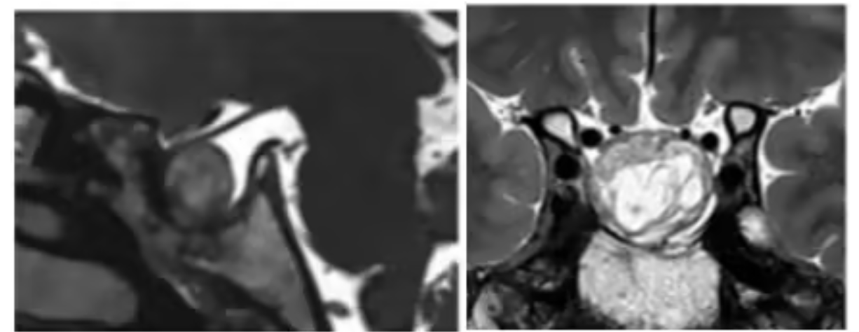
Figure 4: MRI one year after surgery
The importance of brain tumor surgery
The lovely girl Xiaokui had a recurrence 8 years after her first pituitary surgery, and the second surgery still did not achieve complete resection—she even faced blindness. Fortunately, Professor Froelich performed a successful third surgery, and Xiaokui regained her vision. Treatment choices are crucial for brain tumor patients. Every decision during treatment directly affects the prognosis. The initial surgery is particularly important—it must ensure patient safety while maximizing tumor resection. A successful first surgery can avoid many complications and subsequent "compensatory" radiotherapy or chemotherapy. Moreover, a well‑performed initial surgery not only achieves good postoperative results and reduces the patient's physical burden but also lays a solid foundation for subsequent treatment.
How should aggressive pituitary adenomas be treated?
Aggressive pituitary adenomas should be detected early, and a tailored treatment plan should be developed. In most cases, we tend to consider early repeat surgery to maximize safe tumor resection, aiming for long‑term vision preservation in non‑functioning adenomas and biochemical remission in functioning adenomas. Whenever possible, the goal is to preserve normal pituitary function and avoid radiotherapy to reduce long‑term hypopituitarism and radiation exposure. Patients often tolerate repeat surgery better than conventional fractionated radiotherapy. In most non‑functioning adenomas, the recurrence rate is low and the interval relatively long, so surgery can be postponed until a significant recurrence is observed.
Considering the low cure rate of non‑surgical treatment for functioning adenomas (except prolactinomas), repeat surgery offers a better chance of endocrine remission. Residual tumors should be identified by thin‑slice MRI and (functional) PET imaging to define the surgical target, and extended approaches should be used if necessary to achieve remission. If repeat surgery fails or there is no imaging evidence of residual tumor, stereotactic radiosurgery (SRS) can be considered.
In summary, once a brain tumor is found, prompt medical attention is essential. Even if the tumor is in a difficult location with high surgical risks, there is always a suitable surgeon to perform the operation. New therapies and technologies are emerging internationally. Patients should remain confident and do their best to achieve a better quality of life.
References: Sébastien Froelich. Chiasmapexy for secondary empty sella syndrome: diagnostic and therapeutic considerations. Pituitary. 2021 Apr;24(2):292-301.








