2026-07-27
As early as the 1940s, foreign scholars described pituitary adenomas as complex intracranial lesions and reported a postoperative mortality rate of 35% among patients with significant suprasellar extension. Over the past two decades, the World Health Organization (WHO) has updated its classification of pituitary adenomas three times (2004, 2017, 2022 editions). The nomenclature has formally shifted from pituitary adenoma to pituitary neuroendocrine tumors (PitNETs).
A giant pituitary neuroendocrine tumor (G‑PitNET) is defined as a lesion with maximum diameter greater than 4 cm in any dimension, or an estimated tumor volume ≥10 cm³. Giant pituitary tumors account for 5%–15% of all pituitary neoplasms and 0.5% of all intracranial tumors. Typical manifestations include visual impairment, endocrine dysfunction, and neurological compression syndromes caused by mass effect.
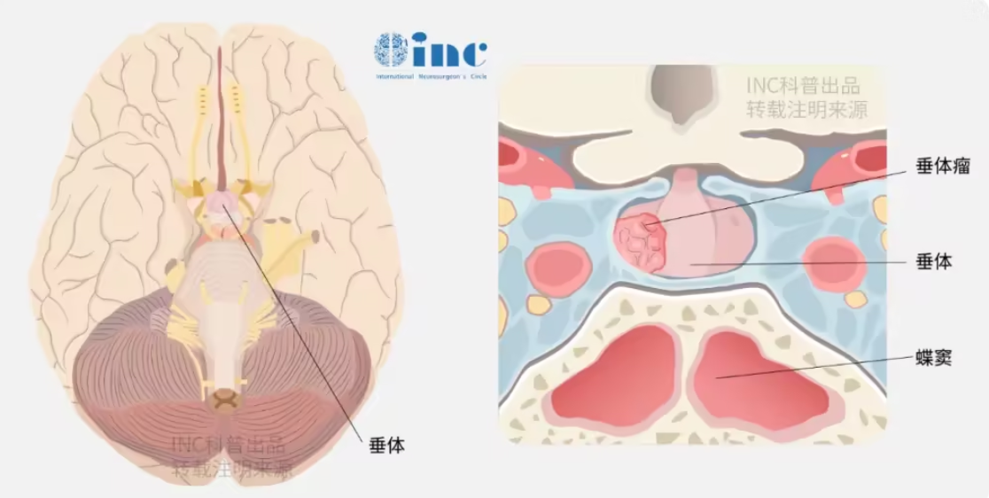
G‑PitNETs remain therapeutically challenging due to their large size, invasiveness and extrasellar extension. The transsphenoidal approach is generally regarded as the gold standard for pituitary tumor surgery, yet critical distinctions exist between operations for giant tumors versus smaller adenomas. Even in specialized tertiary neurosurgical centers, gross total resection (GTR) is achieved in fewer than half of patients after a single procedure, accompanied by substantial surgical morbidity and mortality.

The EANS Consensus Statement
To standardize and optimize surgical management for G‑PitNETs, a task force composed of members of the Skull Base Section of the European Association of Neurosurgical Societies (EANS) and leading international experts developed a consensus guideline:
Surgical management of giant pituitary neuroendocrine tumors: Meta‑analysis and consensus statement on behalf of the EANS skull base section
Professor Sebastien Froelich (France), former Chairman of the Skull Base Surgery Committee of the World Federation of Neurosurgical Societies (WFNS), Chair of Neurosurgery at Lariboisière Hospital in Paris and member of the World Neurosurgery Advisory Group (WANG) under International Neurosurgeon's Circle (INC), was one of the core contributors.
The publication conducted a systematic review and meta‑analysis of articles published between January 2000 and January 2020, enrolling 1,263 patients with G‑PitNETs. The mean tumor diameter was 5.0 ± 0.5 cm. In 50.5% of cases, lesions were Knosp Grade 3 or 4, or confirmed intraoperatively to invade the cavernous sinus. Below is a summary of key data and recommendations.
Key Clinical Data from the Meta‑Analysis
Clinical Presentation
Visual symptoms were the most common clinical presentation (90.1%), followed by endocrine deficits in 57% (Figures 2, 3). Nearly one-third of patients (27%) had complete anterior pituitary insufficiency at diagnosis, whereas diabetes insipidus was rare (2%) (Figure 3). Among these surgical series, 260 patients (260/1222, 21%) had functional tumors, while non-functional tumors were detected in 79% of cases.
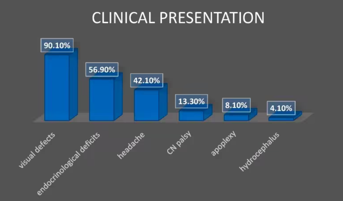
Figure 2: The most common clinical presentation was visual deficits, followed by anterior pituitary hypofunction and headache symptoms.
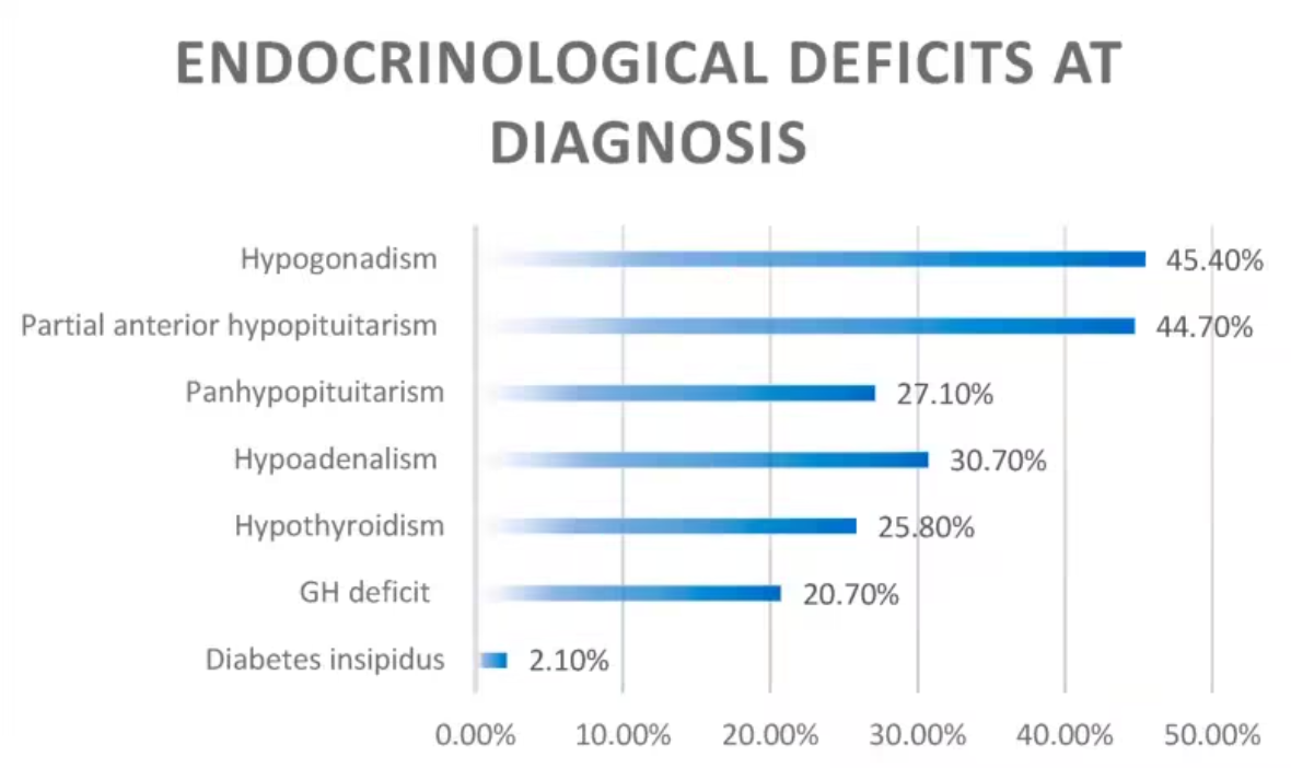
Figure 3: Details the endocrine deficits at diagnosis. The most common finding was hypogonadism, whereas nearly one-third of patients presented with panhypopituitarism. Diabetes insipidus was uncommon at diagnosis.
Surgical techniques
Prof. Froelich and colleagues analyzed the different surgical techniques used in each study, classified as: endoscopic endonasal approach (used in 40.3% of cases), microscopic transsphenoidal approach (34% of cases), transcranial approach (18.7% of cases), and combined approach (7% of cases, using both endonasal and transcranial routes).
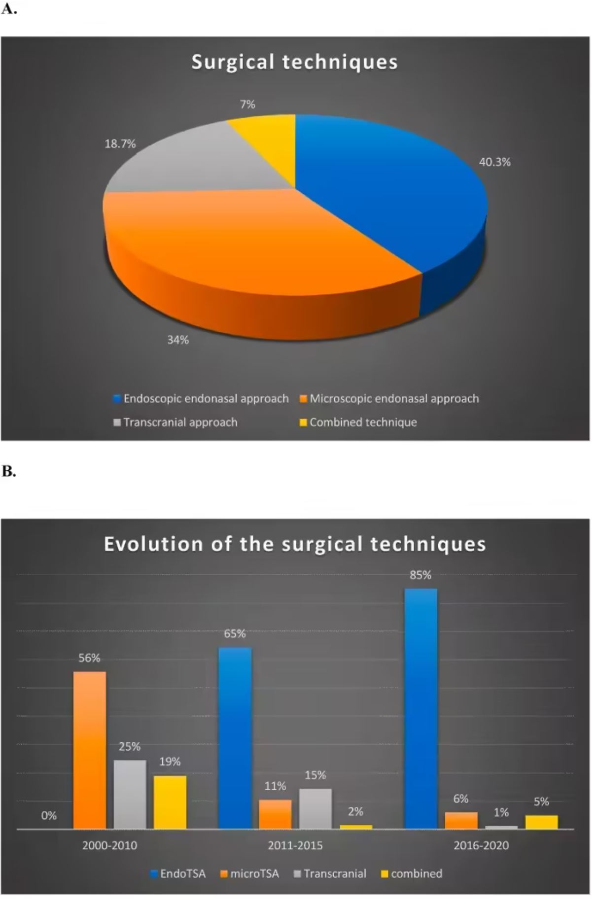
Figure 4A: Summarizes the different surgical techniques used in the papers. Among 1229 patients, 495 (40.3%) underwent an endoscopic endonasal approach (EEA), 418 (34%) underwent a microscopic transsphenoidal approach, 230 (18.7%) underwent a transcranial approach, and 86 (7%) underwent a combined approach (endonasal plus transcranial). Figure 4B: Summarizes the temporal evolution of the different techniques over time.
Among the 403 patients treated with EEA, the meta-analysis showed that 33.8% (±3.9%) achieved gross total resection (GTR), whereas among 185 patients treated via the microscopic transsphenoidal approach, GTR was achieved in 35.8% (±7.9%). In 54 patients treated via the transcranial approach, GTR was achieved in 35.3% (±14.9%), and in 45 patients treated via the combined approach, GTR was achieved in 41.1% (±12.1%).
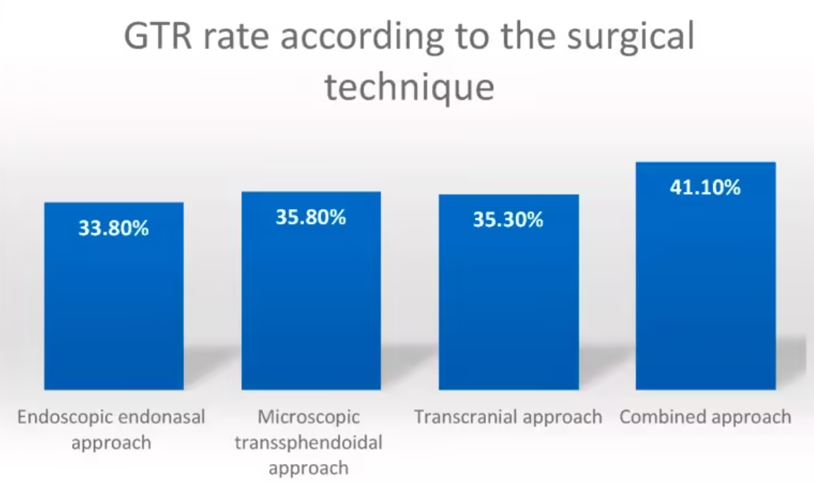
Figure 5: Gross total resection (GTR) rates according to different surgical techniques.
Based on data analysis from the studies, the endocrine improvement rate was 26.1%, with no significant difference among the different surgical approaches. New partial anterior pituitary insufficiency occurred in 21% of patients, and new panhypopituitarism occurred in 2.2%. New permanent diabetes insipidus occurred in 4.9% of cases. Postoperative visual improvement (in terms of visual acuity and visual field recovery) was seen in 75.5% of patients, whereas visual deterioration occurred in 3.1%. The median follow-up was 42.9 months (range 15.5–106.8 months).
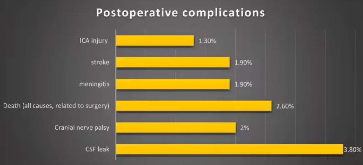
Figure 9 summarizes the most common postoperative complications. The most common complication was cerebrospinal fluid (CSF) leakage, which was rare and reported in less than 4% of cases.
Adjuvant radiotherapy was administered for growing residual or recurrent tumors in 33% of cases, and adjuvant medical therapy was used in 58% of cases to achieve biochemical remission of functional tumors. Tumor recurrence was reported at the last follow-up in 10.4% of cases (CI: 5.9%–14.8%), and reoperation was performed in 6% of cases (3%–9%).
Surgical management
Apart from giant prolactinomas, most GPitNETs require surgical treatment to relieve the compressive effect of the tumor on neural tissues. They are generally associated with limited GTR rates, hence a higher recurrence rate, increased postoperative morbidity and mortality, and an overall poor long-term prognosis. For non-functional GPitNETs, the goal of surgery should be maximal tumor resection to relieve tumor compression on the optic apparatus, pituitary gland, and pituitary stalk, while preserving quality of life. For functional GPitNETs, in addition to relieving optic chiasm compression, the aim is to normalize hormone secretion and, if possible, restore pituitary physiological function. The surgical approach should be tailored according to the size and extent of the GPitNET, its configuration, the need for hormone therapy, and patient-specific treatment goals.
To achieve these goals, several issues should be addressed preoperatively, for example:
1.Is the endonasal approach preferred when managing GPitNETs?
2. What are the indications for transcranial surgery?
3. How should postoperative pituitary apoplexy be managed in GPitNETs?
Is the endonasal approach preferred when managing G-PitNETs?
The endonasal approach is the most commonly used approach for G-PitNETs. In Prof. Froelich’s meta-analysis, which included articles published over the past 20 years, 74.3% of patients underwent an endonasal approach; endoscopic surgery accounted for 40.3% and microscopic transsphenoidal surgery for 34%. No statistically significant difference was found in resection rates between the two techniques. However, over the past decades, several studies have compared endoscopic and microscopic transsphenoidal approaches in the treatment of PitNETs. In terms of resection rates, the endoscopic technique was associated with better outcomes, including visual and endocrine results, particularly for tumors with significant suprasellar and parasellar extension. In 2012, Komotar et al. conducted a meta-analysis including 478 patients with tumors >30 mm and reported a higher rate of GTR in the endoscopic group compared with the microscopic group (P<0.008).
In fact, no statistically significant difference was found in resection rates between endonasal and transcranial approaches. The choice between transsphenoidal and transcranial approaches reported in different series is likely based on individual tumor characteristics to achieve optimal resection. In addition to the surgical approach and adjuncts, factors influencing surgical outcomes include tumor shape, diaphragma sellae opening, multicompartmental extension, lateral intradural extension beyond the internal carotid artery, subarachnoid invasion with encasement of vessels, cavernous sinus invasion, and tumor consistency. Round, dumbbell-shaped, and multilobulated adenomas have each been associated with lower GTR rates. The presence of multiple compartments is an important factor limiting the extent of resection and is often accompanied by arterial encasement and extension into the subarachnoid space.
In addition to the classic transsphenoidal approach, an extended endoscopic endonasal approach may be used when the tumor has marked suprasellar extension and a small sella. Moreover, an extended approach may be considered when the tumor has subfrontal extension.
It is well known that cavernous sinus invasion is a limiting factor for achieving GTR in PitNETs, especially when the lateral compartment of the sinus is involved or the carotid artery is encased. Analysis of the literature on large lesions also confirms this. Tumors within the cavernous sinus can be managed by experienced surgeons via an endoscopic transcavernous approach, particularly for functional tumors, although the efficacy of resecting extensive intracavernous extensions for therapeutic purposes remains controversial.
All these factors should be considered together in a preoperative algorithm (Figure 10), and the main goal is to achieve the safest maximal resection to avoid postoperative complications.
We recommend that the endoscopic endonasal approach be preferred when managing GPitNETs. Depending on the extent, morphology, and anticipated consistency of the lesion, an extended approach may be performed. (Grade C)
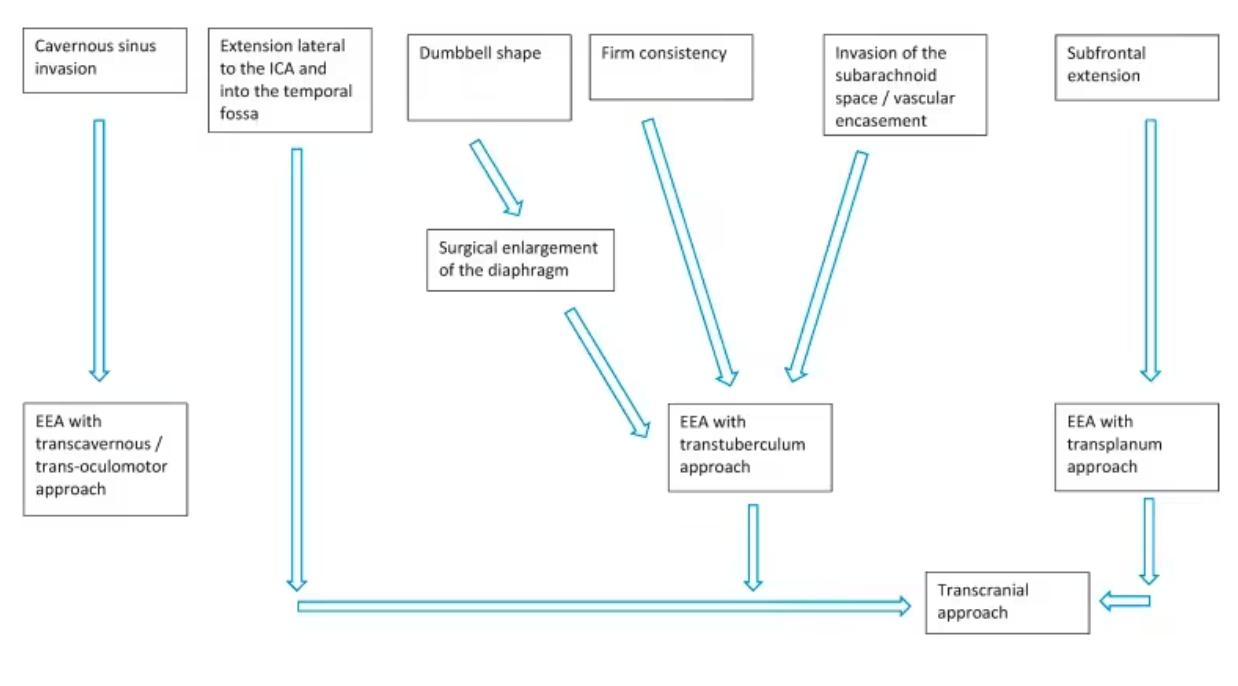
Figure 10: Algorithm for the surgical management of G-PitNETs, based on factors to be considered in selecting the most appropriate surgical approach.
What are the indications for transcranial surgery in giant pituitary tumors?
Since the introduction of endoscopy, increasing experience and technical refinements have allowed the extended endoscopic endonasal approach to be more widely used, even for third-ventricle and subfrontal extensions of pituitary tumors, thereby limiting the use of the transcranial approach to selected cases.
Nevertheless, in some unique and carefully selected cases, transcranial techniques remain appropriate; they may be used as a standalone approach or in combination with a transsphenoidal approach. Flexibility in selecting the optimal surgical method is essential, and the tumor morphology, extent, invasiveness, consistency, and likelihood of surgical cure should be considered.
Currently, the transcranial approach is an option in 0.5–4% of pituitary adenoma cases.
After consensus among the working group members, the indications for the transcranial approach in the treatment of GPitNETs are:
•Tumors with multicompartmental extension that may invade the subarachnoid space and encircle the arteries of the circle of Willis
•Tumors with cisternal encasement of the optic or oculomotor nerves
•Tumors with lateral extension to the supraclinoid internal carotid artery and invasion of the temporal fossa
•Tumors with extensive subfrontal extension
In some cases, if the tumor has expanded through the oculomotor triangle (a common pathway for these tumors to achieve such lateral extension), EEA can be used in experienced hands to manage tumors with lateral extension to the ICA.
In selected cases, a combined endonasal and transcranial approach may be used to combine the advantages of both methods. Depending on the consistency of the lesion, we recommend using the endonasal approach for the bulk of the lesion, while the transcranial approach would address those parts of the tumor that cannot be reached by the extended endoscopic endonasal approach. When the tumor extends into the medial cavernous sinus and enters the basal cisterns with encasement of neurovascular structures, it can be resected via a transcranial skull base approach (transcavernous if needed), which should be tailored according to the extent of tumor extension.
We recommend using a transcranial approach combined with an endonasal approach in selected cases, i.e., for GPitNETs with multicompartmental morphology, subarachnoid invasion with arterial and/or nerve encasement, and ICA lateral extension to the temporal fossa. (Grade C)
Summary of recommendations
• It is recommended to define GPitNETs as tumors with a maximum diameter >40 mm; CT can be used when MRI is contraindicated. (Grade C)
•It is recommended that G-PitNETs with more complex and multilobulated structures be treated in tertiary medical centers, because they are more surgically challenging than more regular round suprasellar G-PitNETs. (Grade C)
•Preoperative endocrine and ophthalmologic evaluation is recommended to determine the patient's preoperative hormonal and visual status. (Grade C)
•It is recommended that the endoscopic endonasal approach be preferred when managing GPitNETs; the approach can be extended according to the extent, morphology, and anticipated consistency of the lesion. (Grade C)
•It is recommended that a transcranial approach combined with an endonasal approach be used in selected cases, such as GPitNETs with multicompartmental morphology, arterial and/or nerve encasement, subarachnoid invasion, and lateral extension of the ICA to the temporal fossa.
•It is recommended that craniotomy be preferred for residual tumor after endonasal surgery in giant tumors, especially for tumors with a firm consistency. In tertiary centers with extensive endoscopic experience, extended endoscopic surgery with extracapsular resection can be performed.
INC International Surgical Professors for Complex Pituitary Tumors

Prof. Bertalanffy (Germany)
Areas of expertise: Surgical treatment of tumors and vascular lesions in complex locations, including the skull base, brainstem, eloquent areas, cerebrum, cerebellum, and spinal cord. Specializes in tumor resection and nerve anastomosis for lesions in complex regions such as the skull base and brainstem. Has performed over a thousand successful brainstem lesion surgeries, achieving gross total resection without damaging neurological function with superb surgical technique.

Prof. Froelich (France)
Areas of expertise: Skull base tumor resection via endoscopic endonasal approach; minimally invasive neuroendoscopic surgery for complex intracranial tumors such as vestibular schwannomas, pituitary adenomas, chordomas, and craniopharyngiomas in challenging locations. His endoscopic “chopstick” technique not only improves the tumor resection rate but also provides better prognostic outcomes for patients.

Prof. William T. Couldwell (USA)
Areas of expertise: Surgical treatment of meningiomas in complex locations such as the petroclival region and posterior fossa, as well as skull base nerve tumors, pituitary tumors, craniopharyngiomas, epilepsy, and cerebrovascular neurosurgery. Has extensive experience with successful meningioma resections and good prognostic outcomes.








