2026-07-20
Pituitary adenomas rank among the most prevalent intracranial neoplasms, second only to gliomas and meningiomas, accounting for 8%–15% of all intracranial tumors. The vast majority are histologically benign, yet a subset of patients experience postoperative tumor recurrence: previously resolved clinical symptoms reappear, follow-up MRI/CT detects regrowth or enlargement of residual tumor tissue, or endocrine testing shows secondary elevation of hormone levels that had normalized after initial resection. Recurrent pituitary adenomas represent a persistent clinical challenge for neurosurgeons, and reoperation carries substantially higher technical difficulty compared to primary resection; standard intraoperative imaging tools alone cannot deliver optimal tumor removal outcomes.
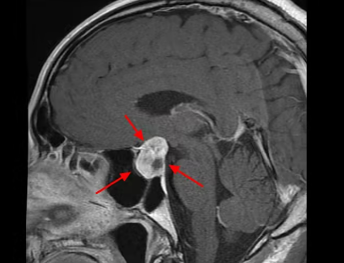
Nowadays, endoscopic endonasal transsphenoidal resection has become the mainstream surgical approach for pituitary adenomas. It avoids multiple complications associated with traditional microscopic transsphenoidal surgery, including severe tissue trauma, nasal septal perforation, nasal cavity infection and mucosal atrophy. Its core advantages include minimal operative trauma, high-definition panoramic visualization, broad anatomical exposure and close-up inspection of lesions, enabling ultra-precise, delicate dissection to reduce surgical injury and complication rates while improving therapeutic efficacy. Patients experience mild postoperative discomfort, shorter hospital stays and lower overall medical costs. High-definition neuroendoscopic transsphenoidal resection for pituitary adenomas and other intracranial masses has become a leading minimally invasive surgical paradigm worldwide.
First reported for clinical use by Dr. Black’s research team at Harvard University in 1996, intraoperative MRI (iMRI) has gained widespread adoption across neurosurgical subspecialties including intracranial tumor resection, functional neurosurgery and stereotactic biopsy, thanks to its unique strengths: real-time intraoperative imaging, automatic correction of brain shift artifacts, and high-precision guidance for dissection and puncture procedures.
For pituitary adenoma surgery—especially resection of invasive or recurrent lesions—iMRI delivers irreplaceable advantages unavailable from conventional preoperative navigation imaging:
1. Maximize radical tumor removal and minimize postoperative residual neoplastic tissue;
2. Rigorously safeguard eloquent brain regions governing speech, motor function and vision;
3. Achieve lesion resection with minimal trauma for pituitary adenomas and gliomas alike;
4. Lower long-term recurrence risk for high-grade gliomas;
5. Reduce iatrogenic injury to critical cranial nerves adjacent to the tumor, cutting short-term and permanent postoperative disability rates;
6. Render surgical and biopsy targets directly visible intraoperatively to drastically boost procedural accuracy.
The core tenet of modern minimally invasive neurosurgery is maximal safe lesion resection with minimal damage to intact neurological tissue. Neuroendoscopes feature a fish-eye wide-angle field of view that eliminates the blind zones inherent to pure microscopic transsphenoidal approaches, streamlining dissection and raising the likelihood of gross total resection. That said, neuroendoscopy carries a steep learning curve requiring rigorous standardized training for consistent operative proficiency, which is critical to elevating complete resection rates for pituitary adenomas.
Frameless stereotactic navigation aids intraoperative localization of lesions by guiding surgical corridor design. Even with navigation assistance, residual tumor remains common after transsphenoidal surgery, particularly for adenomas invading the cavernous sinus. Navigation software can define sellar floor osteotomy boundaries and standardize operative margins for precise tumor dissection, yet the technology bears inherent critical limitations:
Intraoperative brain shift creates major spatial misregistration errors based on preoperatively acquired imaging datasets, leading to misleading anatomical localization;
Minor or significant shifts in the patient’s head position relative to the operating table further degrade navigation accuracy.
Alternative intraoperative imaging modalities such as mobile X-ray, intraoperative ultrasound and intraoperative CT have been trialed for transsphenoidal pituitary surgery, but poor soft-tissue resolution, indistinct sellar anatomy and ionizing radiation exposure prevent widespread clinical uptake.
Intraoperative MRI (iMRI) resolves all the above limitations and serves as the optimal complementary imaging tool for neuroendoscopic pituitary surgery. It enables instant, on-table scanning to dynamically assess the extent of tumor resection and refresh navigation datasets in real time, effectively neutralizing spatial errors caused by brain shift. As an ideal intraoperative adjunct for neurosurgery, iMRI delivers unique value during transsphenoidal resection of recurrent pituitary adenomas:
Superior soft-tissue resolution with crisp visualization of sellar and parasellar anatomical structures;
Zero ionizing radiation, making it safe for repeated real-time scanning throughout the operation;
Direct intra-operative identification of microscopic residual tumor hidden in cavernous sinus recesses, suprasellar cisterns and other blind zones unreachable by endoscope visual inspection alone, allowing immediate secondary resection during the same procedure to minimize leftover tumor tissue.
Multiple international clinical studies have validated the safety and efficacy of iMRI-assisted transsphenoidal pituitary surgery. A retrospective analysis of 73 patients with giant pituitary adenomas recorded a baseline gross total resection rate of 58.9% with standard neuroendoscopy alone; with 3.0T iMRI real-time guidance, the complete resection rate rose significantly to 72.6%. No adverse events or complications related to iMRI equipment use were documented in this cohort.
In summary, iMRI dynamically eliminates navigation drift, visualizes occult residual tumor and expands the scope of safe resection during neuroendoscopic removal of recurrent pituitary adenomas, reliably boosting gross total resection rates while maintaining procedural safety.








