2026-07-20
When skull base tumors such as meningiomas, pituitary adenomas, chordomas and craniopharyngiomas develop inside the brain, surgery is generally the primary treatment option. Yet many patients wonder whether craniotomy is mandatory for these operations. The answer is negative.
Evolution of Neurosurgical Instruments and Techniques
Neurosurgery is a discipline constantly upgrading its surgical tools. The field has advanced from early open neurosurgery to microsurgery, and now minimally invasive endoscopic neurosurgery. Surgical equipment has undergone revolutionary upgrades: from hand-cranked drills to auto-stop craniotomy devices, from single-lens operating microscopes to fluorescence microscopes. Neuroendoscopy has emerged and matured, bringing widespread adoption of minimally invasive skull base procedures that avoid traditional craniotomy. Global neurosurgical technical innovation never stalls.
Neuroendoscopy: Cutting-Edge Therapy for Pituitary Adenomas
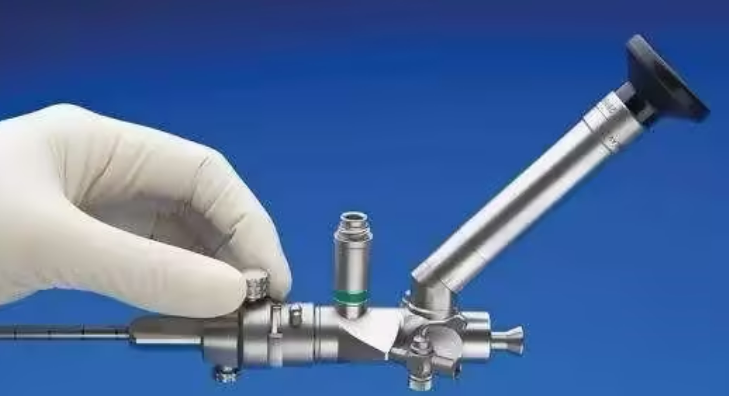
As a flagship minimally invasive neurosurgical technique, neuroendoscopy has expanded its indications across all neurosurgical subspecialties. When combined with an operating microscope, endoscopy delivers unparalleled intraoperative advantages.
A Pair of Sharp Eyes to Visualize All Tumor Tissue
The neuroendoscope acts as a surgeon's high-precision magnifying lens. It can pass through narrow natural surgical corridors to reach the tumor surface, offering multi-magnification, multi-angle panoramic visualization of the operative field. Even tiny tumor remnants hidden in blind anatomical recesses can be clearly identified for complete removal.
Endoscopic surgery features minimal soft tissue trauma and reliable hemostasis. The native bone flap can be preserved, eliminating the need for secondary cranioplasty to repair skull defects after open surgery.
Skull base lesions including meningiomas, pituitary adenomas, chordomas and craniopharyngiomas can achieve complete resection via advanced endoscopic techniques.
For pituitary adenomas specifically, most lesions are resectable through the endoscopic endonasal transsphenoidal corridor. All manipulations are performed under direct endoscopic visualization, outperforming pure microsurgery in surgical smoothness, gross total resection rate and minimal tissue injury. Nasal packing is unnecessary postoperatively, greatly reducing patient discomfort.
Beyond skull base tumors, neuroendoscopic equipment also enables effective treatment of hydrocephalus, intraventricular cysts, intracranial arachnoid cysts, chronic subdural hematoma, intraventricular hemorrhage and cerebral hemorrhage.
Representative Case: Endoscopic Resection of Pituitary Adenoma
A 37-year-old French female patient was diagnosed with a 9 mm pituitary microadenoma centered within the sella turcica. She underwent minimally invasive endoscopic endonasal transsphenoidal single-nostril resection. The surgeon adopted the world-famous endoscopic “chopstick technique” to achieve en bloc gross total resection of the entire tumor. The patient received standardized adjuvant endocrine therapy postoperatively. Three years of follow-up confirmed no tumor recurrence and zero surgical complications.
This endoscopic pituitary adenoma resection was performed by Professor Sebastien Froelich, a preeminent pioneer of neuroendoscopic skull base surgery widely recognized as a leading authority in skull base neurosurgery. He is a core member of the World Neurosurgery Advisory Group (WANG) under INC International Neurosurgical Physicians Group, and one of the youngest specialists ever appointed Chairman of the Skull Base Surgery Committee of the World Federation of Neurosurgical Societies (WFNS).
Neuroendoscopy originated in Western countries and has only seen widespread clinical development over the past two decades. Most advanced endoscopic surgical systems are imported, and complete hardware suites are only available at a small number of top-tier tertiary hospitals in China. Surgical outcomes featuring such high gross total resection rates and favorable long-term prognosis remain uncommon domestically.
Professor Froelich practices at Hôpital Lariboisière, a historic Paris university hospital equipped with world-leading neurosurgical infrastructure. Cases achieving near-perfect radical tumor resection are routine here, supported by state-of-the-art intraoperative MRI and real-time neurophysiological monitoring systems to guarantee surgical safety and efficacy.
“During endoscopic procedures, we utilize cutting-edge technologies including 3D endoscopes paired with 4K ultra-high-definition imaging to optimize endoscopic visualization quality. To maximize resection completeness and surgical outcomes, we integrate microscopes, endoscopes and exoscopes within a single operation,” Professor Froelich explained in an exclusive interview with INC. The hospital's integrated endoscopic surgical platform ranks among the most advanced worldwide.
A Global Master of Endoscopic Skull Base Neurosurgery
The skull base is a labyrinthine anatomical corridor densely populated with cranial nerves and critical cerebral arteries. Safe, complete endoscopic tumor removal demands comprehensive, precise anatomical knowledge of the skull base, paired with exceptional proficiency in endoscopic instrumentation.
Few neurosurgeons worldwide possess deeper expertise in endoscopic skull base surgery than Professor Sebastien Froelich, Chair of Neurosurgery at Hôpital Lariboisière in Paris.
Milestones of Professor Froelich's Technical Innovation
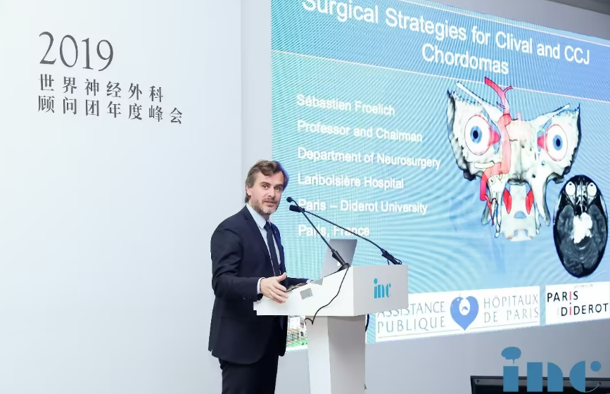
As early as 2010, Professor Froelich's team pioneered endoscopic single-nostril transsphenoidal clipping for anterior communicating artery aneurysms, a groundbreaking achievement in neuroendovascular and endoscopic neurosurgery. His team subsequently invented the dual-hand “chopstick technique” to resolve coordination challenges during endoscopic dissection. Renowned for his extraordinary dexterity, sharp clinical insight and landmark contributions to endoscopic skull base surgery, he is regularly invited worldwide to deliver hands-on technical training workshops.
He has traveled to China multiple times for academic exchange: at the 2019 WFNS Special Congress and International Endoscopic Neurosurgery Symposium held at Xuanwu Hospital (Capital Medical University), the Suzhou Neurosurgery Salon at the First Affiliated Hospital of Soochow University, and the annual summit of INC's World Neurosurgery Advisory Group in Shanghai. Professor Froelich generously shares his innovative surgical techniques and clinical experience with Chinese neurosurgeons to drive collective clinical progress, earning widespread respect among domestic neurosurgical practitioners for his modest, collaborative








