2026-07-20
Pituitary adenomas are common extra-axial intracranial tumors, accounting for 10%–15% of all intracranial neoplasms and 33%–35% of sellar lesions. They arise from the adenohypophysis and are histologically benign. Nevertheless, a small subset grows invasively, exhibiting biological behaviors resembling malignant lesions; these are termed invasive pituitary adenomas.
Such tumors invade adjacent anatomical structures, making complete surgical resection difficult and leading to a high postoperative recurrence rate. They can occur at any age, most commonly in patients aged 30 to 60 years, rarely in those over 60 or under 10, with no obvious gender predominance. Pituitary adenomas are classified by biological behavior into non-invasive and invasive subtypes. First proposed by Jefferson in 1940, invasive pituitary adenomas are defined as lesions whose tumor cells infiltrate and damage surrounding normal tissues including dura mater, bone, brain parenchyma and paranasal sinuses. Advances in imaging technology have enabled extensive research on the radiological characteristics of these tumors.
Most pituitary adenomas follow an indolent clinical course with slow recurrence, their natural history limited by anatomical barriers to radical surgical excision. Certain subtypes, however, feature rapid regrowth and treatment resistance. Many specialized centers regard a Ki-67 index of 7%–10% as a clinically meaningful biomarker for aggressive biological activity. Under current pathological classification standards, Ki-67 remains a core marker of invasiveness, though no universal diagnostic cutoff value has been established. Radiological evidence of cavernous sinus or clival invasion is also recognized as a clinical indicator of aggressive tumor behavior.
Part 2: Standard Treatment Strategies for Invasive Pituitary Adenomas
Clinical guidelines for recurrent and invasive pituitary adenomas have been issued across North America and Europe, with a shared core recommendation that these complex lesions be managed via multidisciplinary team (MDT) care delivered by pituitary subspecialist neurosurgeons.
1. Guidance from Congress of Neurological Surgeons (CNS)
Based on Level 2 and Level 3 clinical evidence, the guidelines recommend early adjuvant radiotherapy (conventional fractionated radiotherapy CRT or stereotactic radiosurgery SRS) for postoperative residual tumor to lower the risk of future progression. The panel acknowledges limited supporting evidence and notes that optimal timing for postoperative radiotherapy requires further clinical research.
The guidelines identify repeat surgical resection as the primary intervention for recurrent or residual adenomas, reserving CRT/SRS for patients unfit for reoperation due to high surgical risk.
2. European Society of Endocrinology (ESE) Guidelines for Aggressive Pituitary Tumors
The ESE guidelines also recommend expert repeat surgery as first-line intervention prior to alternative adjuvant therapies. Adjuvant radiotherapy is only advised for postoperative residual lesions when clear clinical markers of aggressiveness are present (e.g., radiological invasion or elevated Ki-67 index).
Temozolomide chemotherapy is recommended for progressive aggressive pituitary adenomas or pituitary carcinomas with documented tumor growth, albeit supported by low-quality clinical evidence. No clear consensus specifies the optimal disease stage to initiate temozolomide treatment.
While both sets of guidelines are limited by weak supporting evidence, they establish a standardized framework for routine clinical practice.
3. Comprehensive Clinical Treatment Logic
Aggressive invasive pituitary adenomas require early detection and individualized treatment regimens. Clinicians generally prioritize early repeat surgery to achieve maximal safe tumor resection:
For non-functional adenomas: Long-term visual function preservation is the primary goal;
For functional adenomas: Biochemical remission of excess hormone secretion is targeted.
Where feasible, surgeons aim to preserve intact native pituitary function and avoid radiotherapy, reducing the long-term risk of permanent hypopituitarism and cumulative radiation exposure. Patients tolerate repeat surgical intervention far better than prolonged fractionated radiotherapy. For most non-functional adenomas with low recurrence risk and long disease-free intervals, reoperation can be delayed until radiological progression is clearly documented.
With the exception of prolactinomas, medical monotherapy achieves low biochemical remission rates for most functional adenomas, making repeat surgery the superior option to normalize hormone levels. Thin-slice MRI and functional PET imaging precisely delineate residual tumor to define surgical targets; extended transsphenoidal corridors are adopted as needed to achieve radical resection.
If repeat surgery fails to reduce tumor burden or no residual lesion is visible on imaging, stereotactic radiosurgery (SRS) is indicated.
Invasive pituitary adenomas typically demand multimodal combined therapy and repeated salvage interventions. Temozolomide chemotherapy is increasingly deployed earlier in the disease trajectory, yet surgery remains irreplaceable for debulking mass lesions and safeguarding neurological function, despite the technical challenges of repeat pituitary operations.
Part 3: Clinical Case Managed by Professor Sebastien Froelich (INC France)
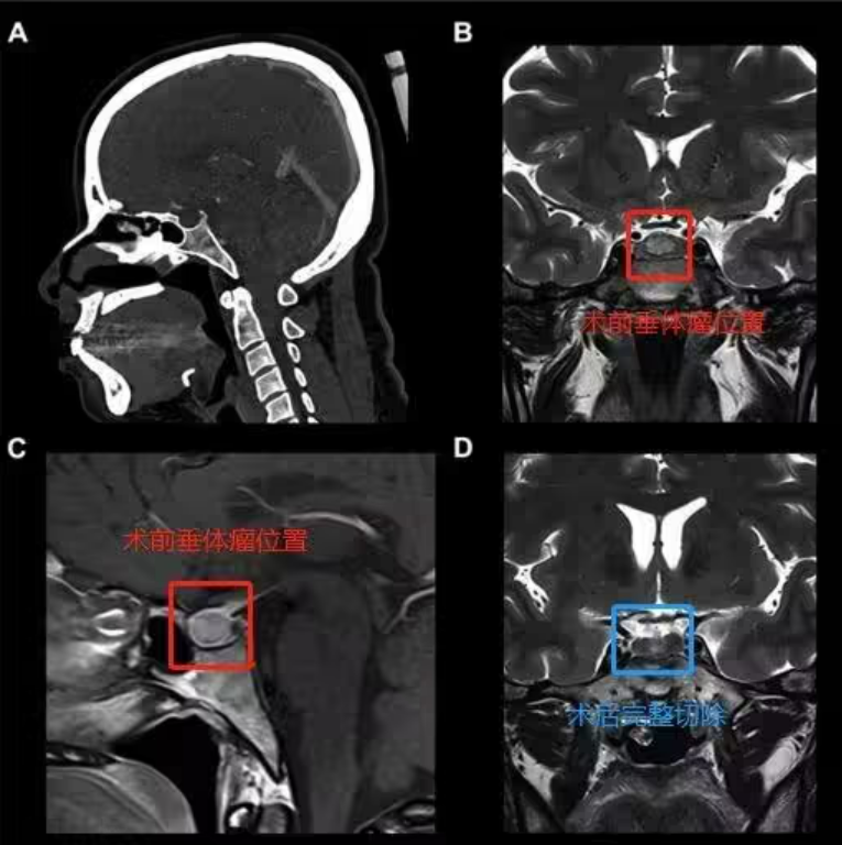
A 37-year-old French female patient was diagnosed with a 9 mm central sellar pituitary microadenoma. She underwent minimally invasive endoscopic endonasal transsphenoidal resection using the internationally renowned endoscopic “chopstick technique” pioneered by Professor Sebastien Froelich, enabling en bloc gross total resection of the lesion. The patient received standardized adjuvant endocrine therapy postoperatively, with no tumor recurrence and zero surgical complications recorded over three years of follow-up.
Preoperative imaging (red marker): Axial and coronal MRI show a 9 mm microadenoma centered within the sella turcica; CT confirms sellar periosteal pneumatization of the sphenoid sinus.
Intraoperative procedure: The chopstick technique enables wide sphenoidotomy, meticulous hemostasis, sharp dissection along the pseudocapsular plane, and en bloc tumor excision (piecemeal resection is avoided to eliminate residual neoplastic tissue).
Postoperative imaging (blue marker): Gross total resection is confirmed on direct intraoperative visualization, followed by full-course endocrine regulation treatment.
Brief Introduction to Professor Sebastien Froelich
Professor Sebastien Froelich of INC International Neurosurgical Physicians Group, former Chairman of the Skull Base Surgery Committee of the World Federation of Neurosurgical Societies (WFNS), specializes in minimally invasive endoscopic endonasal resection of skull base tumors, including complex lesions such as pituitary adenomas, chordomas and craniopharyngiomas. His innovative endoscopic chopstick dual-hand technique substantially elevates gross total resection rates and improves long-term functional outcomes for patients with skull base neoplasms.








