2026-07-17
Over the past two decades, the World Health Organization (WHO) has released three iterations of pituitary adenoma classification (2004, 2017, 2022). The nomenclature has officially shifted from pituitary adenomas to pituitary neuroendocrine tumors (PitNETs). A giant pituitary neuroendocrine tumor (G-PitNET) is defined as a lesion with a maximum diameter exceeding 4 cm in any plane, or a total tumor volume ≥10 cm³. Giant pituitary tumors account for 5%–15% of all pituitary neoplasms and 0.5% of all intracranial tumors. Their typical clinical manifestations include visual dysfunction, endocrine impairment, and compressive neurological syndromes caused by mass effect.
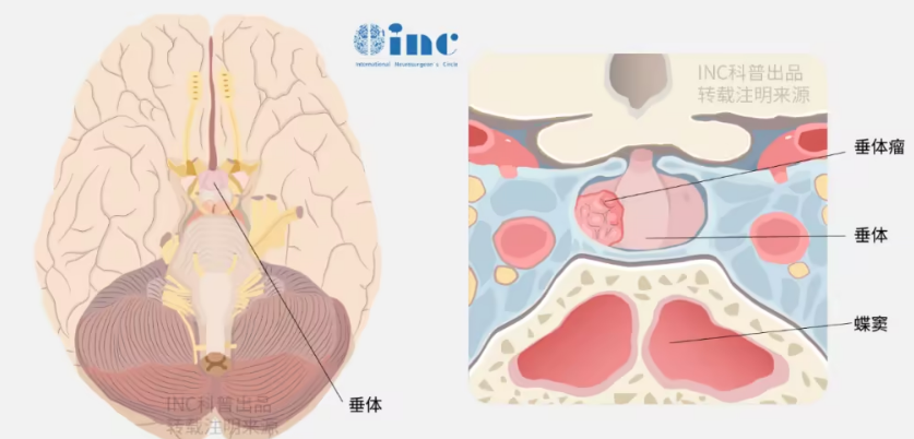
Giant PitNETs remain a major therapeutic challenge due to their large size, invasive biological behavior and extensive parasellar/suprasellar extension. The endonasal transsphenoidal approach is widely recognized as the gold standard for pituitary tumor resection, yet surgery for giant lesions differs substantially from operations on smaller adenomas. Even at specialized tertiary care centers, gross total resection (GTR) is achieved in fewer than half of patients after a single operation, accompanied by considerable perioperative morbidity and mortality risks.
To standardize and optimize the surgical management of G-PitNETs, a task force consisting of members of the Skull Base Section of the European Association of Neurosurgical Societies (EANS) and leading international specialists developed a formal consensus statement with clinical recommendations. Professor Sebastien Froelich, member of the World Neurosurgery Advisory Group (WANG) under INC International Neurosurgical Physicians Group and former Chairman of the Skull Base Surgery Committee of the World Federation of Neurosurgical Societies (WFNS), was a core participant in this consensus development.
The consensus research, titled Surgical management of giant pituitary neuroendocrine tumors: Meta-analysis and consensus statement on behalf of the EANS skull base section, was published in the journal Brain and Spine.
The paper conducted a systematic literature review and meta-analysis covering studies published between January 2000 and January 2020, enrolling a total of 1,263 patients with G-PitNETs. The mean tumor diameter was 5.0 ± 0.5 cm. In 50.5% of cases, lesions were classified as Knosp grade 3–4 or confirmed intraoperatively to invade the cavernous sinus. Below is a full summary of the core consensus conclusions and clinical recommendations for G-PitNET treatment.
Core Consensus Summary from the EANS Skull Base Section
1. Clinical History, Physical Examination and Endocrine Workup
Visual deficits constituted the most prevalent presenting symptom (90.1%), followed by endocrine dysfunction (57%). Approximately one-third (27%) of patients presented with complete anterior hypopituitarism at initial diagnosis, while diabetes insipidus was extremely rare (only 2%). Among the cohort of 1,222 evaluable patients, 260 (21%) harbored functional PitNETs, and 79% had non-functional adenomas.
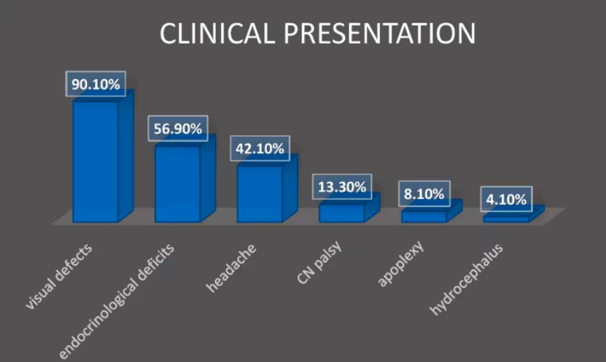
Figure 2: The most common clinical manifestations are visual deficits, followed by anterior hypopituitarism and headache.
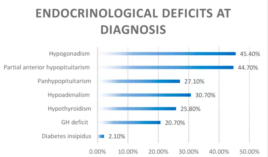
Figure 3: Breakdown of endocrine deficits at diagnosis. Hypogonadism is the most frequent finding; nearly one-third of patients develop panhypopituitarism, whereas diabetes insipidus is seldom detected preoperatively.
2. Surgical Modalities and Resection Outcomes
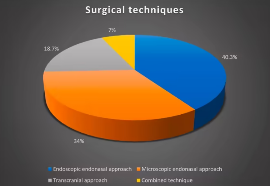
Professor Froelich’s team categorized all operative techniques recorded in included studies into four groups: endoscopic endonasal approach (EEA, used in 40.3% of cases), microscopic transsphenoidal approach (34%), transcranial approach (18.7%), and combined endonasal-transcranial approach (7%).
A.
B.
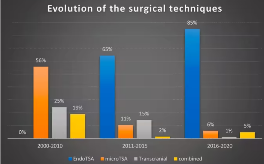
Figure 4A: Distribution of surgical techniques across 1,229 patients: 495 (40.3%) endoscopic endonasal surgeries, 418 (34%) microscopic transsphenoidal surgeries, 230 (18.7%) transcranial surgeries, and 86 (7%) combined dual approaches. Figure 4B illustrates the chronological shift in technique utilization over decades.
Meta-analyzed gross total resection (GTR) rates by surgical corridor:
• Endoscopic endonasal approach (403 patients): 33.8% ± 3.9% GTR
• Microscopic transsphenoidal approach (185 patients): 35.8% ± 7.9% GTR
• Transcranial approach (54 patients): 35.3% ± 14.9% GTR
• Combined dual approach (45 patients): 41.1% ± 12.1% GTR
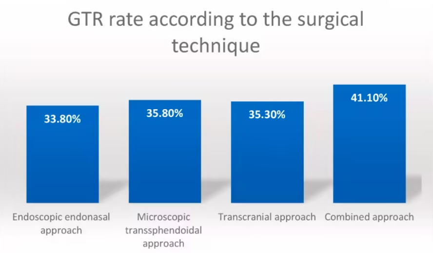
Figure 5: Comparative GTR rates across all four surgical modalities.
Key perioperative and long-term functional outcomes from pooled data:
• Endocrine improvement rate: 26.1%, with no statistically significant difference across surgical techniques.
• New partial anterior hypopituitarism: 21% of patients; new complete panhypopituitarism: 2.2%.
• New permanent diabetes insipidus: 4.9%.
• Visual improvement (acuity + visual field recovery): 75.5%; permanent visual deterioration: 3.1%.
• Median clinical follow-up: 42.9 months (range: 15.5–106.8 months).
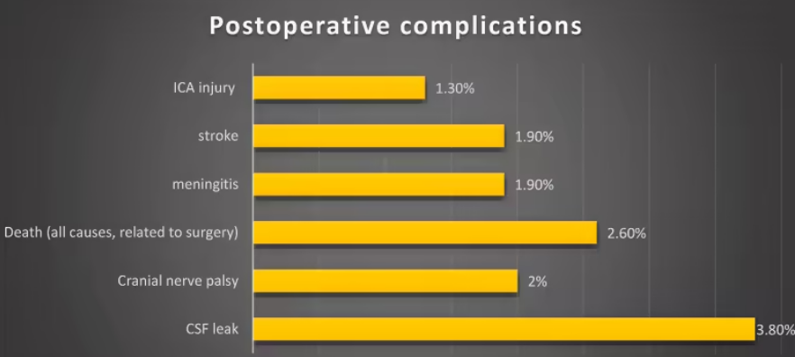
Figure 9: Overview of postoperative complications. CSF rhinorrhea was the most common adverse event, occurring in less than 4% of all cases.
Adjuvant therapy and recurrence statistics:
• Adjuvant radiotherapy was administered to 33% of patients with progressive residual or recurrent tumor.
• Adjuvant medical therapy was used in 58% of functional PitNET cases to achieve biochemical remission.
• Overall tumor recurrence rate at final follow-up: 10.4% (95% CI: 5.9%–14.8%).
• Reoperation rate: 6% (95% CI: 3%–9%).
3. General Surgical Principles
With the exception of giant prolactinomas suitable for primary medical management, surgical decompression is indicated for nearly all G-PitNETs to relieve mass effect on intracranial neural structures. These lesions are associated with low single-stage GTR rates, high recurrence risk, elevated perioperative morbidity/mortality and suboptimal long-term prognosis.
Surgical goals for non-functional G-PitNETs: Maximize safe tumor resection to decompress the optic apparatus, pituitary gland and pituitary stalk while preserving patients’ quality of life.
Surgical goals for functional G-PitNETs: In addition to visual pathway decompression, normalize excess hormone secretion and restore intact pituitary endocrine function whenever feasible.
Surgical corridor selection must be individualized based on tumor size, extension pattern, tissue consistency, need for postoperative medical therapy and patient-specific functional targets.
Four core preoperative clinical questions addressed by the consensus panel:
• Is the endonasal approach the first-line option for G-PitNET resection?
• What are formal indications for transcranial surgery?
• How should postoperative pituitary apoplexy be managed?
• Is the endonasal approach preferred for G-PitNETs?
The endonasal corridor represents the most widely utilized surgical route for giant PitNETs. Across the 20-year meta-analysis cohort, 74.3% of patients underwent endonasal surgery (40.3% endoscopic, 34% microscopic). No statistically significant difference in overall GTR rates was observed between endoscopic and microscopic transsphenoidal techniques.
Multiple comparative studies have demonstrated superior visual and endocrine outcomes with endoscopic approaches for tumors with extensive suprasellar and parasellar extension. A 2012 meta-analysis by Komotar et al. including 478 patients with tumors >30 mm confirmed significantly higher GTR rates in the endoscopic cohort (P < 0.008).
No statistically significant difference in resection rates exists between transsphenoidal and transcranial approaches; corridor selection in individual clinical series is tailored to unique tumor morphology to maximize resection extent. Independent of surgical corridor and intraoperative adjuncts, factors limiting complete resection include tumor shape, size of sellar diaphragm opening, multicompartmental extension, lateral intradural extension beyond the internal carotid artery (ICA), subarachnoid invasion with arterial encasement, cavernous sinus infiltration and tumor consistency. Round, dumbbell-shaped and multilobulated adenomas all correlate with reduced GTR likelihood. Multicompartmental extension is a major limiting factor, frequently accompanied by subarachnoid tumor extension wrapping intracranial arteries.
Beyond standard transsphenoidal corridors, extended endoscopic endonasal approaches are indicated for lesions with prominent suprasellar extension and small native sella turcica, as well as tumors with significant subfrontal extension. Cavernous sinus invasion is a well-documented barrier to GTR, especially with lateral cavernous sinus extension or complete ICA encasement, a finding validated by pooled analysis of giant tumor cohorts. Experienced surgeons can resect intrasinus tumor components via endoscopic transcavernous corridors for functional adenomas, though the clinical benefit of aggressive resection of extensive cavernous sinus tumor remains debated.
All anatomical variables must be integrated into preoperative decision-making algorithms (Figure 10), with the primary objective of maximal safe resection to avoid avoidable postoperative complications.
Consensus Recommendation (Grade C): The endoscopic endonasal approach is the first-line surgical corridor for G-PitNET resection; extended variants may be utilized based on lesion extension, morphology and anticipated tumor consistency.
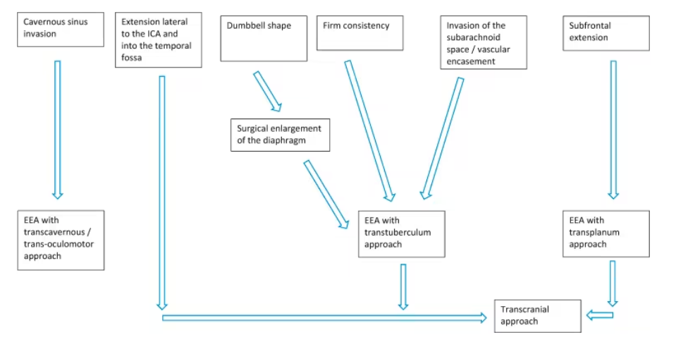
Figure 10: Decision algorithm for G-PitNET surgical management outlining anatomical variables guiding corridor selection.
4. Formal Indications for Transcranial Surgery
Widespread adoption of endoscopy, accumulated surgical experience and technical refinements have expanded the applicability of extended endoscopic endonasal corridors even for tumors extending to the third ventricle and subfrontal region, restricting transcranial surgery to highly selected complex cases. Transcranial approaches may be used alone or combined with endonasal routes, with flexible corridor selection critical to match tumor anatomy, invasiveness, consistency and curative potential. Transcranial surgery accounts for only 0.5%–4% of all pituitary adenoma operations at present.
After panel consensus voting, absolute indications for transcranial resection of G-PitNETs are defined as follows:
• Multicompartmental tumor extension with subarachnoid invasion and encasement of arteries of the circle of Willis;
• Tumors encasing the optic nerve or oculomotor nerve within basal cisterns;
• Lateral tumor extension superior to the clinoid process involving the temporal fossa with ICA encasement;
Massive subfrontal tumor extension.
In select cases with lateral ICA extension confined to the oculomotor trigone (the most common pathway for lateral parasellar tumor spread), experienced surgeons may achieve adequate resection via the endoscopic endonasal approach (EEA). For highly complex lesions, combined endonasal-transcranial surgery leverages the strengths of both corridors: the endonasal route debulks the main intrasellar mass, while transcranial access targets tumor extensions unreachable by endoscopy. Lesions extending medially into the cavernous sinus, basal cisterns and wrapping neurovascular structures can be removed via tailored transcranial skull base corridors (including transcavernous variants as needed).
Consensus Recommendation (Grade C): Combined endonasal-transcranial surgery is recommended for selected G-PitNETs with multicompartmental morphology, subarachnoid invasion with arterial/cranial nerve encasement, and lateral ICA extension into the temporal fossa.
Full List of Grade C Consensus Recommendations
G-PitNETs are defined as lesions with a maximum diameter >40 mm; CT may substitute MRI for patients with absolute MRI contraindications.
Complex multilobulated G-PitNETs should be managed exclusively at tertiary care neurosurgical centers, as they carry substantially higher technical difficulty compared to regular rounded suprasellar giant adenomas.
Complete preoperative endocrine and ophthalmological assessment is mandatory to establish baseline hormonal function and visual status prior to surgery.
The endoscopic endonasal approach is recommended as first-line surgery for G-PitNETs, with extended endonasal variants deployed according to tumor extension, morphology and anticipated tissue consistency.
Combined endonasal-transcranial surgery is indicated for complex multicompartmental G-PitNETs with arterial/cranial nerve encasement, subarachnoid invasion and lateral ICA extension into the temporal fossa.
For residual tumor after endonasal resection of giant adenomas, transcranial surgery is preferred, particularly for firm, fibrotic lesions. At high-volume tertiary centers with advanced endoscopic expertise, extended extradural endoscopic resection may be attempted as an alternative.








