2026-07-17
Within the confined sellar compartment spanning merely around 5.5 square centimeters lies a high-stakes anatomical battlefield critical to visual and neurological function. Fifty-nine-year-old Anna faced a severe clinical crisis: a sellar pituitary cytoma compressed her pituitary stalk and optic chiasm like an immense boulder, triggering progressive visual deterioration. The lesion's intimate proximity to vital blood vessels and cranial nerves drastically elevated surgical hazards. Nevertheless, guided by the personalized surgical strategy devised by Professor Sebastien Froelich of the INC International Neurosurgical Physicians Advisory Group, this highly complex operation achieved safe tumor resection with complete preservation of all vital neurovascular structures, paving the way for the patient's full recovery.
Rare Tumor Behind Rapid Vision Loss: Clinical Dilemmas of Sellar Pituitary Cytomas
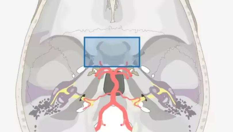
The sellar region acts as a core neuroendocrine hub housing the pituitary gland, optic nerves, optic chiasm, internal carotid arteries and other vital structures, with only a 3-centimeter narrow operative corridor available. The slightest iatrogenic trauma can lead to permanent irreversible functional deficits. Anna suffered marked visual decline over three months, and subsequent imaging confirmed a large sellar pituitary cytoma. Classified as a rare benign glial neoplasm of WHO Grade I, this tumor predominantly arises within the sellar and suprasellar spaces. Slow mass expansion commonly induces endocrine dysfunction, visual impairment and persistent headache; severe cases develop anterior hypopituitarism manifesting as amenorrhea, sexual dysfunction and diabetes insipidus, or obstructive hydrocephalus.
Treatment of this lesion is hindered by multiple inherent challenges:
1. Anatomical complexity: The tumor lies adjacent to the pituitary stalk, optic chiasm and perforating blood vessels, requiring ultra-careful dissection to avoid neurovascular injury.
2. Dense tissue adhesions: Firm adherence to the hypothalamus and brainstem raises intraoperative hemorrhage risk and the likelihood of permanent neurological deficits.
3. Hypervascularity: Rich intratumoral vasculature creates substantial difficulties in controlling intraoperative bleeding.
4. Diagnostic limitations: The tumor is easily missed on preliminary workup, often only definitively identified via intraoperative pathological consultation, amplifying complication risks.
Individualized Surgical Strategy: Resolving Dual Major Barriers of Sellar Tumor Resection
As a leading skull base neurosurgeon and member of INC's global neurosurgery advisory panel, Professor Froelich formulated a tailored operative plan targeted specifically at Anna's unique tumor anatomy.
Challenge 1: Balancing Deep Tumor Adhesions with Functional Preservation
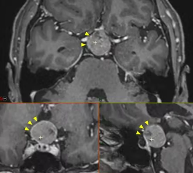
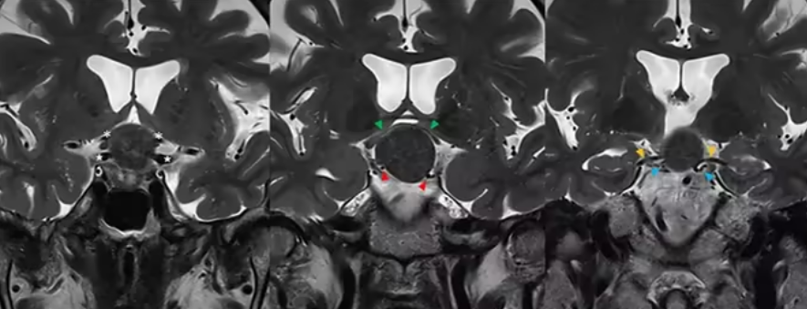
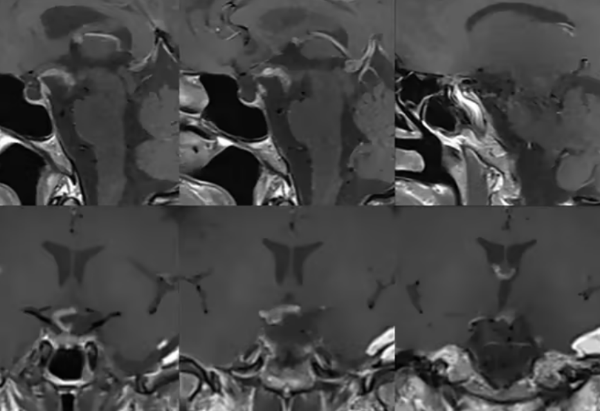
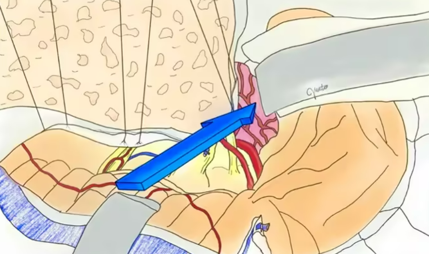
MRI demonstrated a spherical mass occupying the posterior pituitary infundibulum and suprasellar cistern, compressing and thinning the optic chiasm with dense adhesions to the internal carotid arteries and anterior cerebral arteries. Professor Froelich abandoned the standard endoscopic endonasal approach (EEA) in favor of the posterior petrosal corridor. This route delivers multi-angle surgical exposure from beneath the temporal lobe, lowers the incidence of postoperative cerebrospinal fluid rhinorrhea, and is uniquely suited to preserve the right pituitary stalk and intact pituitary endocrine function. Guided by the core principle of prioritizing long-term functional prognosis over radical resection, blind pursuit of gross total removal was avoided to safeguard irreplaceable neurovascular tissue.
Challenge 2: Securing Hemostasis and Intraoperative Safety
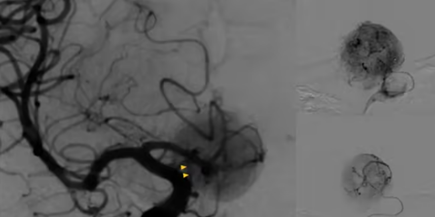
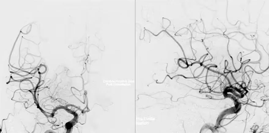
Given the tumor's robust blood supply, preoperative digital subtraction angiography (DSA) pinpointed the left superior hypophyseal artery as the primary feeding vessel. Professor Froelich implemented preoperative endovascular embolization to occlude the proximal segments of dural feeding branches. Post-embolization angiography confirmed occlusion of approximately 90% of the tumor's blood supply, drastically reducing anticipated intraoperative blood loss.
Precision Dissection for Optimized Prognosis: Key Milestones from Operation to Rehabilitation
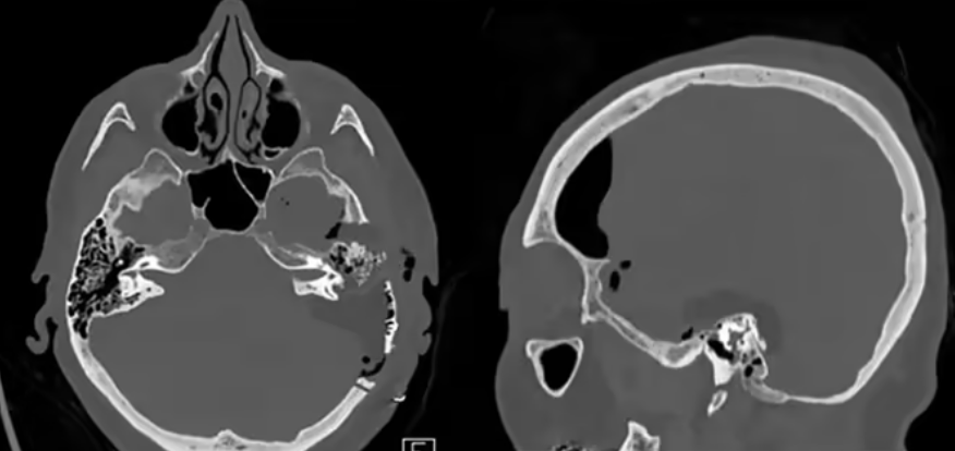
Under high-power surgical microscopy, Professor Froelich meticulously dissected the tumor away from surrounding critical structures. To safeguard endocrine function, a thin layer of tumor tissue was intentionally preserved over the pituitary stalk and optic chiasm surface. Thanks to effective preoperative embolization, the procedure proceeded without hemorrhage or intraoperative adverse events, and the bulk of the tumor was successfully excised. Postoperative histopathology confirmed the diagnosis of pituitary cytoma. Anna was discharged after a seven-day hospital stay with no new neurological or endocrine deficits. Six-month follow-up imaging revealed no progression of the residual tumor, accompanied by substantial visual improvement and restored quality of life.
Suitability Analysis of the Posterior Petrosal Approach for Sellar Pituitary Cytomas
Professor Froelich's published research notes that while the posterior petrosal corridor was initially developed for craniopharyngioma resection, it delivers distinct advantages for pituitary cytoma management:
Direct visualized dissection: Access behind the pituitary stalk enables clear delineation of the tumor's interface with perforating vessels, significantly improving rates of intact pituitary function preservation.
Mitigated risk profiles: Compared with the endoscopic endonasal approach (EEA), this corridor minimizes trauma to hypothalamic feeding arteries, lowering the risk of postoperative diabetes insipidus and hypothyroidism.
This approach nevertheless carries inherent limitations, including elevated risk of injury to cranial nerves III and IV, alongside a steep technical learning curve. Professor Froelich emphasizes that a full-cycle customized regimen integrating preoperative embolization, personalized corridor selection and targeted functional protection must be designed preoperatively based on tumor location, vascular supply and individual patient characteristics, striking an optimal balance between maximal safe resection and functional preservation.
Brief Introduction to Professor Sebastien Froelich
A world-renowned pioneer in endoscopic skull base neurosurgery, Professor Froelich boasts extensive clinical experience treating complex lesions including chordomas, pituitary adenomas and craniopharyngiomas. His innovative “chopstick technique” and dual-modality combined endoscope-microscope surgical framework have consistently boosted gross total resection rates and long-term patient functional outcomes. In recent years, Professor Froelich has repeatedly traveled to China for academic lectures and live demonstrative surgeries, delivering novel individualized treatment solutions to numerous patients with sellar lesions.
Treatment for sellar pituitary cytomas is far more than a straightforward mission to eliminate tumor tissue—it constitutes a critical defense of intact neurological function. Professor Froelich's personalized treatment paradigm verifies that rigorous preoperative stratification, innovative surgical corridor design and meticulous intraoperative dissection enable safe, effective tumor control within high-risk sellar anatomy, providing an invaluable clinical reference for precision care of rare skull base neoplasms.








