2026-07-17
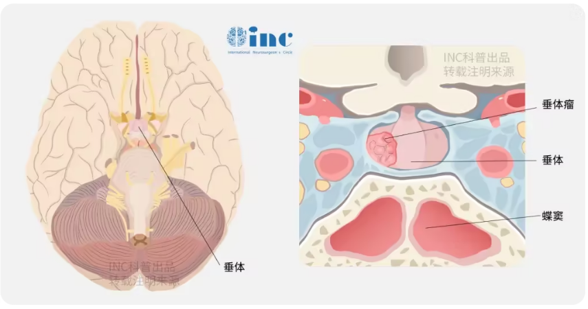
Deep at the base of the brain lies the pituitary gland, a pea-sized vital endocrine organ less than one centimeter across. It serves as the core regulator of the human hormonal balance, precisely sustaining the orderly operation of all physiological processes. When the pituitary gland is invaded by a tumor, its normal function is disrupted. The mass not only compresses surrounding structures such as the optic nerves and blood vessels but also triggers systemic disorders through abnormal hormone secretion or disrupted normal endocrine regulation, presenting symptoms including menstrual irregularities, acromegaly, osteoporosis and thyroid dysfunction. Particularly for macroadenomas with a diameter exceeding 4 centimeters, a far more complex “endocrine storm” may be induced.
I. Clinical Characteristics and Epidemiological Data of Pituitary Tumors
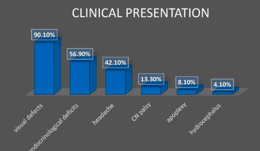
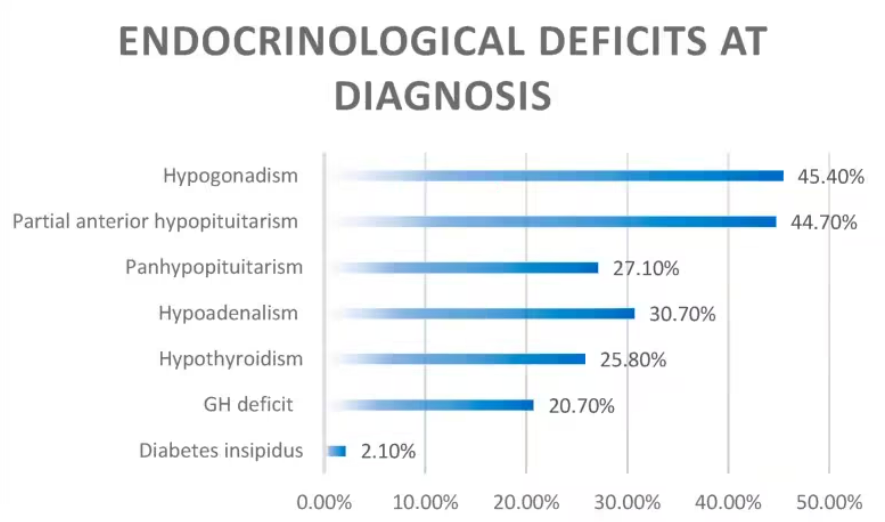
Pituitary adenomas originate from pituitary tissue and the vast majority are benign, accounting for approximately 15% of all intracranial neoplasms. Giant pituitary macroadenomas are defined as lesions with a maximum diameter ≥4 cm or a volume ≥10 cm³, primarily manifesting as visual disturbances, endocrine dysfunction and compressive neurological symptoms. Clinical statistics reveal that 90.1% of patients present with visual deficits as their initial complaint, 57% have endocrine abnormalities, and complete anterior hypopituitarism is already present in 27% of patients at the time of diagnosis, while diabetes insipidus is rare with an incidence of only 2%.
Such tumors impair pituitary function either through autonomous hormone overproduction or mass compressive effects. Functional adenomas (e.g., prolactinomas, growth hormone-secreting adenomas) produce clinical manifestations tied to excess hormone secretion. Non-functional adenomas mainly cause visual deterioration and hypopituitarism by compressing the optic chiasm, pituitary stalk or hypothalamus.
Endocrine deficits are commonly detected upon diagnosis. Hypogonadism is the most prevalent finding, and nearly one-third of patients develop panhypopituitarism, whereas diabetes insipidus is seldom seen at initial presentation.
II. Therapeutic Strategies and Key Surgical Principles for Pituitary Macroadenomas
(1) Surgical Indications and Core Treatment Goals
Except for giant prolactinomas, where medical therapy may be attempted as first-line management, most pituitary macroadenomas require surgical intervention with the following primary objectives:
For non-functioning adenomas: Maximize tumor resection to decompress the optic nerves, pituitary gland and pituitary stalk, and restore neurological function.
For functional adenomas: Achieve adequate decompression while striving to recover normal hormonal secretion and physiological pituitary function.
(2) Surgical Corridor Selection and Technical Advances
With advances in neuroendoscopic techniques, endoscopic endonasal transsphenoidal surgery has become the dominant therapeutic modality. This approach reaches the sellar region via the natural nasal passage, featuring minimal tissue trauma and clear panoramic visualization that enables comprehensive inspection of sellar anatomy, lowering risks of residual tumor and iatrogenic injury. Surgical plans are customized based on individual tumor morphology:
Staged Resection Protocol
Prioritize resection of the intrasellar basal vascular core of the tumor to reduce intraoperative hemorrhage.
Dissect parasellar extensions with extreme caution to preserve neurovascular structures within the cavernous sinus.
Fully decompress suprasellar residual lesions to avoid traction injury to the visual pathway.
Key Determinants of Surgical Outcomes
Tumor consistency: Hard, fibrotic or recurrent lesions carry lower gross total resection expectations to prevent excessive aggressive dissection.
Degree of invasion: The scope of subarachnoid infiltration dictates the safe surgical resection boundary.
Growth pattern: Midline vertically extending tumors are well suited for endoscopic transsphenoidal surgery; eccentric lateral extension requires adjusted operative strategies.
III. Surgical Risks and Complication Management
(1) Common Postoperative Complications
Cerebrospinal fluid (CSF) rhinorrhea: Incidence ranges from 0.5% to 4%. Initial leaks can be managed conservatively with lumbar cistern drainage to facilitate spontaneous closure; refractory cases require secondary reconstructive surgery to prevent secondary meningitis.
Internal carotid artery injury: Usually caused by off-midline dissection. Precise vascular localization intraoperatively reduces the risk of arterial laceration.
Optic nerve injury: May result from mechanical traction or postoperative intrasellar hematoma. Intraoperative neuroprotection and close serial visual monitoring postoperatively are mandatory.
(2) Clinical Solutions Proposed by International Leading Experts
Professor Henry W.S. Schroeder, a top specialist affiliated with INC International Neurosurgical Physicians Group, adopts endoscope-assisted microsurgery for large pituitary adenomas invading the cavernous sinus, designing individualized regimens tailored to each tumor's growth pattern.
Representative Case Illustration
For a giant pituitary adenoma with suprasellar extension and cavernous sinus invasion, staged endoscopic endonasal transsphenoidal resection was performed. Autologous abdominal fat grafts were used to reconstruct the sellar floor intraoperatively to prevent CSF leakage.
Technical Advantages
Nasal packing is eliminated to maintain unobstructed nasal breathing after surgery. Postoperative MRI confirms complete gross total resection with satisfactory preservation of native pituitary endocrine function.
Endoscopic endonasal transsphenoidal resection has become the mainstream treatment for pituitary adenomas.

IV. Long-Term Postoperative Surveillance and Prognosis
Individualized follow-up schedules are formulated based on postoperative pathological reports and serial hormonal testing:
Patients with functional adenomas require regular hormonal surveillance, with adjuvant medical therapy or radiotherapy administered if indicated.
Patients with non-functional adenomas undergo periodic sellar MRI to screen for tumor recurrence and assess anatomical recovery of sellar structures.
Clinical research confirms that early surgical intervention significantly improves visual acuity and endocrine function in patients. Customized surgical planning and neuroprotective techniques are critical to minimizing complications and elevating long-term quality of life.
Conclusion
Treatment for pituitary tumors, especially giant macroadenomas, demands dual therapeutic priorities: radical tumor resection and preservation of intact endocrine and neurological function. Leveraging innovative endoscopic technology and personalized multimodal strategies, international neurosurgical experts achieve safe gross total resection and optimized long-term prognoses even for highly complex lesions. Patients experiencing progressive visual loss or persistent hormonal disturbances are advised to undergo timely radiological and endocrine testing to secure optimal therapeutic timing.








