2026-07-17
The pituitary gland sits within the sellar region at the center of the skull base, surrounded by vital structures including the optic nerves, internal carotid arteries, cavernous sinuses and hypothalamus. The optic nerves govern visual function; compression from a pituitary adenoma can trigger visual field defects and, in severe cases, complete irreversible blindness
56-year-old Mr. Rudiger encountered a life-threatening health crisis. Initially suffering from declining visual acuity and restricted peripheral vision, he sought care at an ophthalmology clinic yet received no definitive diagnosis. A subsequent neurosurgical consultation uncovered the true culprit: a large pituitary adenoma.
Mr. Rudiger's adenoa presented highly complex anatomical extension. The tumor grew upward along the midline and laterally invaded the right temporal lobe. Its expansion exceeded the standard working corridor of the endoscopic endonasal transsphenoidal approach. Treatment options were limited to craniotomy or extended transsphenoidal corridors, both of which carried substantial risk of severe iatrogenic injury with any minor intraoperative misstep. The surgery demanded maximal preservation of intact neurological function alongside maximal radical tumor resection, drastically elevating operative difficulty.
Overwhelmed with anxiety, Mr. Rudiger harbored numerous concerns: Could the tumor be removed safely? What represented the optimal surgical strategy? Was gross total resection achievable? Would permanent postoperative complications arise? Professor Henry W.S. Schroeder, former Chairman of the Endoscopy Committee of the World Federation of Neurosurgical Societies (WFNS) and member of the World Neurosurgery Advisory Group (WANG) under INC International Neurosurgical Physicians Group, delivered a reassuring verdict: surgery was feasible and could be performed in a minimally invasive fashion.
As Professor Schroeder predicted, the operation proceeded uneventfully. The procedure achieved complete tumor resection while fully preserving native normal pituitary parenchyma, and follow-up imaging confirmed nearly invisible postoperative scarring.
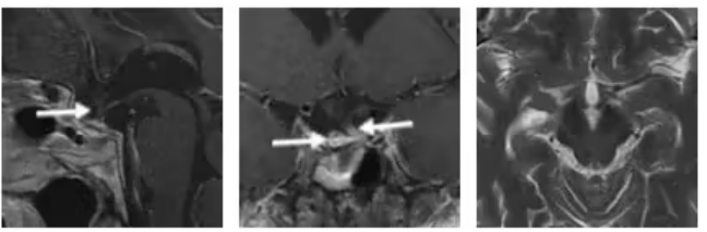
Gross Total Resection Achieved with Intact Normal Pituitary Tissue Preserved
Tumor Compressing the Optic Chiasm with Temporal Lobe Extension Creates Severe Surgical Challenges
The pituitary gland resides in the central sellar compartment of the skull base, encircled by irreplaceable vital anatomy: optic nerves, internal carotid arteries, cavernous sinuses and the hypothalamus. Compression of the optic nerves by adenomas results in visual field deficits and potential blindness. The internal carotid arteries serve as the brain's primary blood supply; accidental intraoperative injury may precipitate catastrophic hemorrhage or cerebral infarction. The cavernous sinus houses arteries and cranial nerves, and tumor invasion here induces extraocular movement dysfunction and facial hypoesthesia. The hypothalamus acts as the body's central hormonal regulatory center; damage can trigger lifelong refractory endocrine dysfunction.
Mr. Rudiger's visual deterioration confirmed direct mass effect of the adenoma upon his optic chiasm. Surgeons were required to dissect tumor tissue within the extremely confined sellar space—a technically demanding maneuver where the slightest error could induce permanent visual loss or irreversible hypothalamic dysfunction.
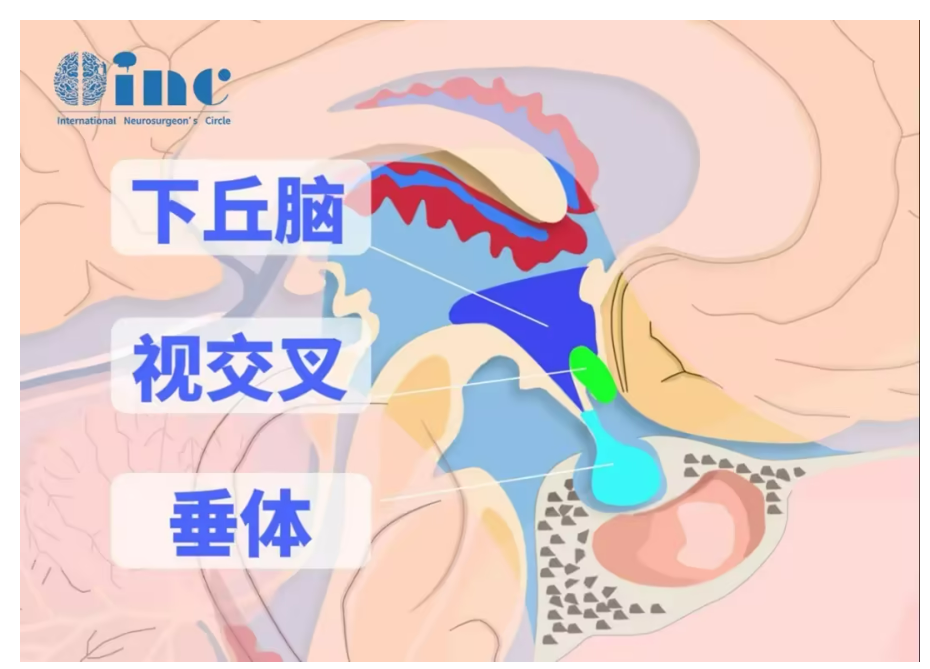
Any minute intraoperative misstep risks irreversible visual impairment or permanent hypothalamic dysfunction.
Compounding the complexity, the adenoma extended laterally into the right temporal lobe, frequently accompanied by dural infiltration or firm adhesions to native brain parenchyma. Conventional curettage techniques inevitably leave residual tumor cells behind. Given the risk of cavernous sinus and temporal lobe parenchymal invasion, dissection demanded extreme precision; minor mishaps could trigger massive hemorrhage or permanent neurological deficits, further amplifying operative risk and complexity.
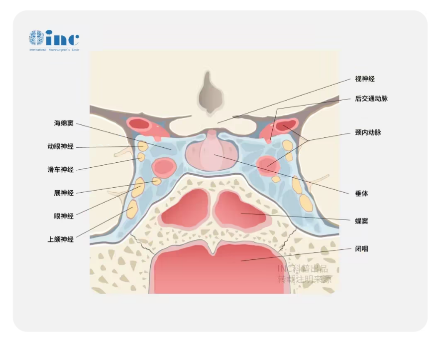
Any trivial intraoperative error may lead to life-threatening hemorrhage or permanent neurological damage.
Endoscopic endonasal transsphenoidal surgery is a widely adopted minimally invasive technique, yet it cannot achieve complete resection for lesions with temporal lobe extension such as Mr. Rudiger's adenoma, due to limited distal reach of endoscopes. The alternative open craniotomy approach—subfrontal or pterional corridor—targets suprasellar and temporal lobe tumor components but carries significant soft tissue trauma, with potential temporal lobe injury that may precipitate postoperative epilepsy or cognitive impairment. Against this clinical dilemma, careful selection of a tailored surgical plan became critical.
Combined Staged Surgery with Supraorbital Keyhole Corridor Delivers Minimally Invasive Resection
Complete radical resection via a single surgical corridor was deemed nearly impossible for this complex case. Drawing upon decades of extensive clinical experience, Professor Schroeder performed thorough radiological and anatomical evaluation and finalized a two-stage combined-corridor surgical strategy.
Stage one consisted of endoscopic endonasal resection of intrasellar and midline tumor components. Stage two employed open craniotomy focused on resecting the right parasellar, temporal lobe-extending tumor bulk. When designing the protocol, Professor Schroeder balanced maximal tumor removal against single-procedure operative trauma, selecting the supraorbital keyhole craniotomy as the open surgical corridor to eradicate all tumor tissue while minimizing iatrogenic injury.
Intraoperative visualization revealed characteristic gray-white normal pituitary tissue with a soft, readily dissectable consistency.
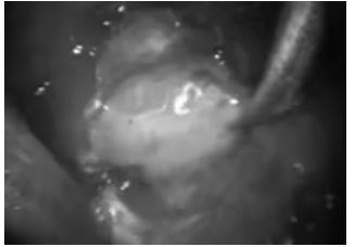
The “keyhole” concept in cranial surgery holds small yet pivotal clinical significance, described figuratively via the term “keyhole bone window”. Keyhole craniotomy does not prioritize arbitrarily tiny incisions for cosmetic purposes alone; incision and bone flap dimensions are individualized based on lesion anatomy, adhering to the core principle of “sufficient exposure for lesion resection with minimal cranial bony trauma”.
During Mr. Rudiger's first operation, Professor Schroeder performed endoscopic endonasal transsphenoidal resection through the natural nasal corridor. Utilizing the panoramic wide-angle visualization of the neuroendoscope, combined with vacuum suction devices and surgical curettes, he sequentially debulked and excised tumor tissue. Intraoperative views clearly delineated soft gray-white intact pituitary parenchyma that separated easily from neoplastic tissue. This first stage achieved complete resection of midline sellar and suprasellar tumor components, yet anatomical constraints left residual adenoma on the right parasellar side.
During the initial postoperative recovery phase, Mr. Rudiger's visual dysfunction improved markedly, confirming effective decompression of the visual pathway with favorable prognostic signals and laying a solid foundation for planning the second-stage operation.
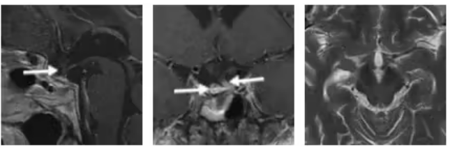
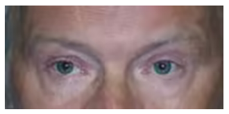
Mr. Rudiger's visual deficits had significantly alleviated.
Two months following the initial procedure, Professor Schroeder performed the second-stage resection via a right supraorbital eyebrow incision craniotomy. He deployed combined dual-modality microscopy and neuroendoscopy: the operating microscope delivered high-definition stereoscopic visualization, while the angled neuroendoscope penetrated blind operative corners. This dual-modality technique minimized retraction of healthy brain tissue and enabled precise dissection of densely adherent tumor parenchyma. Intraoperative inspection demonstrated yellow-hued tumor tissue tightly adhered to adjacent cranial nerves and blood vessels.
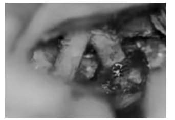
The dual-modality combined technique reduces cerebral retraction and enables precise dissection of adherent tumor tissue.
Postoperative contrast-enhanced MRI verified gross total resection with zero residual tumor; the pituitary stalk and a small volume of adjacent intact pituitary tissue were fully preserved. Six months after surgery, the scar at Mr. Rudiger's right eyebrow was barely perceptible, delivering excellent cosmetic outcomes with which the patient expressed full satisfaction.