2026-07-16
Professor Henry W.S. Schroeder, a distinguished member of the World Neurosurgery Advisory Group under INC International Neurosurgical Physicians Group, states: “The diagnosis and treatment of invasive pituitary adenomas require a departure from conventional mindsets. Every step, from precise preoperative assessment to customized surgical strategy, directly shapes patients' long-term quality of life.”
Li Ming, a 38-year-old male patient, sought medical care for persistent headache and blurred vision and was subsequently diagnosed with an invasive pituitary adenoma. Dubbed “intracranial stubborn lesions”, these tumors are defined by aggressive invasive growth and extremely high recurrence rates, leaving traditional treatment modalities trapped in a vicious cycle of incomplete resection and repeated relapse. As Professor Schroeder emphasizes, breakthroughs across the full clinical workflow are indispensable to improve patient outcomes.
I. Invasive Pituitary Adenomas: Benign Tumors with Malignant-Like Biological Behavior
Core Distinguishing Features That Separate Them from Ordinary Pituitary Adenomas
Unlike slow, circumscribed benign pituitary adenomas, invasive pituitary adenomas act as aggressive intracranial invaders with three hallmark malignant traits.
1. Aggressive Biological Behavior
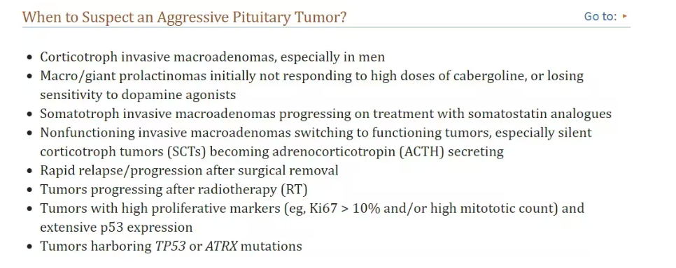
Tumor cells exhibit vigorous proliferative activity, with Ki-67 proliferation index consistently exceeding 10% (normal pituitary tissue registers Ki-67 < 3%). Pathogenic mutations including TP53 and ATRX drive rapid tumor expansion, with an annual growth rate greater than 5 mm. Many subtypes overproduce functional hormones such as prolactin (causing amenorrhea and galactorrhea) or growth hormone (triggering acromegaly), while roughly 30% of invasive prolactinomas develop complete drug resistance to cabergoline.
2. Destructive Anatomical Infiltration
Superior extension compresses the optic chiasm and induces bitemporal hemianopia; lateral invasion penetrates the cavernous sinus and encases more than half the diameter of the internal carotid artery, a definitive imaging marker of invasiveness; inferior erosion destroys bony walls of the sphenoid sinus, raising the risk of spontaneous cerebrospinal fluid (CSF) rhinorrhea. On MRI, these lesions display heterogeneous mixed signals from intratumoral hemorrhage and necrosis, with irregular “moth-eaten” heterogeneous contrast enhancement and ill-defined tumor-parenchyma borders resembling blurred ink wash diffusion.
3. Clinically Refractory Nature
Traditional surgical approaches only achieve gross total resection (GTR) in 30%–50% of cases, accompanied by a 5-year postoperative recurrence rate of 60%–80%, in stark contrast to the under 15% recurrence rate of non-invasive pituitary adenomas. These tumors show limited radiosensitivity, and adjuvant radiotherapy frequently causes permanent hypopituitarism requiring lifelong hormone replacement therapy.
Warning Signs for Early Recognition of Invasive Pituitary Adenomas
Li Ming' s clinical journey illustrates the insidious progression of these aggressive lesions, with key red flags listed below:
• Progressive worsening headache: Transient dull ache evolves into unremitting severe cephalalgia that awakens patients from sleep, triggered by dural stretch from expanding sellar mass.
• Acute visual deterioration: Unilateral or bilateral blurry vision, formal perimetry confirming bilateral peripheral visual field loss secondary to optic chiasm compression.
• Severe endocrine dysfunction: Gynecomastia in males, irregular menses in females, progressive enlargement of hands and feet (classic for GH-secreting invasive adenomas).
• Catastrophic acute presentation: Pituitary apoplexy (intratumoral hemorrhage) induces projectile vomiting and altered mental status, mandating emergent decompressive surgery.
• Definitive Dual Modality Diagnosis: Combined Radiology and Histopathology
Radiological Evaluation
CT imaging reveals expanded sella turcica and destructive erosion of clival bone; Li Ming' s CT scan demonstrated honeycomb-like bony destruction of the sphenoid sinus wall.
Dynamic contrast-enhanced MRI identifies heterogeneous tumor enhancement and cavernous sinus invasion, defined as encasement of over 50% of the internal carotid artery lumen.
Histopathological Confirmation
Tissue specimens obtained via surgical resection or stereotactic biopsy confirm invasive disease per the 2021 WHO Classification of Central Nervous System Tumors when Ki-67 index >15%, p53 immunohistochemical positivity, and histological evidence of vascular invasion are concurrently identified.
II. Limitations of Conventional Treatment Modalities
Three Major Surgical Barriers: Operating Within a Narrow, Vital Cranial Cavity
• The sellar region is densely populated with irreplaceable delicate structures: optic nerves (less than 2 mm in diameter), 4 mm internal carotid arteries, and the hypothalamus. Resection is analogous to intricate embroidery inside a walnut shell, hindered by three unavoidable challenges.
• Severe spatial constraints Conventional operating microscopes offer a limited field of view, failing to visualize blind zones including the medial cavernous sinus wall and floor of the third ventricle. Approximately 20% of residual tumor tissue persists in these hard-to-reach regions (Neurosurgery, 2024).
• Ill-defined tumor margins Invasive adenomas lack a complete fibrous capsule, creating ambiguous boundaries between neoplastic cells and healthy pituitary parenchyma. Aggressive over-resection carries a 5%–10% risk of permanent diabetes insipidus.
• High intraoperative hemorrhage risk Fragile aberrant tumor neovasculature predisposes to uncontrolled bleeding; conventional hemostatic techniques risk irreversible neurovascular injury, forcing premature surgical termination. During Li Ming' s initial operation, massive intraoperative hemorrhage limited tumor resection to only 60% of the lesion volume.
Limited Adjuvant Value of Pharmacotherapy and Radiotherapy
Medical management: Only viable for select prolactinomas; 30% of invasive subtypes develop complete drug resistance. Li Ming received cabergoline for three months yet documented 20% tumor volume enlargement on serial imaging.
Radiotherapy: Reserved for postoperative residual or recurrent lesions, with a therapeutic latency of 1–2 years. Radiation carries a roughly 5% risk of permanent optic nerve injury and cannot reverse pre-existing compressive neurological deficits.
Vicious Recurrence Cycle: Cumulative Risks of Repeated Operations
Patients with subtotal primary resection develop densely adherent recurrent tumors that adhere firmly to adjacent neurovascular structures. The GTR rate drops below 20% during reoperation, while the risk of permanent cranial nerve injury triples. Professor Schroeder stresses: “Primary surgery represents the single best opportunity to achieve cure; treatment delivered by a high-volume, experienced neurosurgical team is non-negotiable.”
III. Breakthrough Solutions: Professor Schroeder' s Precision Integrated Diagnosis & Treatment Framework
Revolutionary Surgical Philosophy: Shift Focus from Tumor Resection to Functional Preservation
Professor Schroeder' s team pioneered a three-dimensional precision surgical protocol tailored to the unique anatomical characteristics of invasive pituitary adenomas.
1. Individualized surgical corridor selection
• Endoscopic endonasal transsphenoidal approach: Indicated for intrasellar and suprasellar lesions, utilizing the natural nasal anatomical corridor to eliminate the morbidity of craniotomy.
• Modified transpterygoid corridor: Designed for cavernous sinus-invasive adenomas; partial resection of the pterygoid bone expands the surgical working space to 0.8–1.2 cm.
2. Real-time multimodal intraoperative monitoring
• Neurophysiological monitoring: Continuous visual evoked potential (VEP) tracking delivers automated alerts when signal attenuation exceeds 10%, preventing irreversible visual loss.
• Indocyanine green (ICG) fluorescence angiography: Intravascular contrast clearly demarcates tumor margins, restricting resection error to less than 0.5 mm.
3. Layered skull base reconstruction A three-layer repair technique combining autologous fat graft, fascia lata, and artificial dura mater reduces postoperative CSF leak rates from the historical 15% down to 3%.
Clinical Case: Successful Gross Total Resection of Recurrent Invasive GH Adenoma
A 45-year-old female patient presented with three prior surgical recurrences of an invasive GH-secreting adenoma fully encasing the internal carotid artery. Professor Schroeder implemented a staged combined endoscopic-microscopic resection strategy:
• Stage 1: 0° endoscope resects intrasellar tumor bulk to reduce intracranial pressure and fully expose cavernous sinus invasive components.
• Stage 2: 30° angled endoscope accesses the cavernous sinus cavity; 8W laser fiber vaporizes microscopic residual tumor while maintaining complete internal carotid artery integrity.
• Stage 3: Adjuvant proton beam radiotherapy targets microscopic residual disease postoperatively. Postoperative MRI confirmed complete gross total resection. The patient' s serum GH level normalized from an initial 45 ng/mL, with no new permanent neurological deficits.
Standardized Postoperative Surveillance to Block Recurrence
Customized long-term follow-up protocols are established for every patient:
• 3 months postoperatively: Contrast-enhanced sellar MRI + full pituitary endocrine panel to evaluate resection extent and residual pituitary function (TSH, morning cortisol, gonadal hormones).
• Starting at 2 years post-surgery: Bi-annual surveillance with Ki-67 biomarker testing; rising proliferation index triggers early interventional therapy.
Stratified recurrence management strategies:
• Recurrent lesion <2 cm in maximum diameter: Single-fraction Gamma Knife radiosurgery with 12–15 Gy marginal dose for long-term tumor growth control.
• Recurrent mass >3 cm or acute optic nerve compression: Reoperation combining cavitational ultrasonic surgical aspirator (CUSA) and full endoscopic exploration to debulk tumor core and minimize traction-related neurovascular trauma.
IV. Cutting-Edge Novel Technologies Reshaping Treatment Standards
1. Molecular Targeted Therapy
Novel small-molecule inhibitor MIRA-101 activates apoptotic pathways in TP53-mutant invasive adenomas, inducing greater than 30% tumor volume reduction in a subset of patients, per Phase II clinical trial data published in Nature Reviews Drug Discovery (2025).
2. Robotic-Assisted Pituitary Surgery
The Da Vinci Xi Surgical System' s 7-degree articulated robotic arms enable ultra-precise 0.2 mm dissection within the narrow cavernous sinus space. Clinical data from Germany' s INI Hospital (2024, 50 consecutive patients) demonstrates a 40% reduction in cranial nerve injury risk compared to standard endoscopic techniques.
3. Combined Immunotherapy Regimens
Dual immune checkpoint blockade (PD-1 inhibitor + CTLA-4 antibody) achieves an objective response rate of 25% in pituitary carcinoma, the malignant end-stage subtype of invasive pituitary adenomas, offering new therapeutic options for drug-refractory advanced disease (Journal of Clinical Oncology, 2024).
V. Patient Clinical Decision-Making Guide
Three Critical Pitfalls to Avoid at Initial Diagnosis
1. Misconception: Mild symptoms equal benign tumor Many invasive adenomas only present with mild headache at early stages; high-resolution sellar MRI is mandatory to rule out aggressive invasion.
2. Overreliance on pharmacotherapy For lesions compressing the optic apparatus, urgent surgical decompression takes priority over prolonged medical observation, as drug therapy cannot reverse established visual nerve damage.
3. Underestimation of primary surgery' s significance The extent of resection during the first operation directly determines long-term prognosis; patients must select neurosurgical centers performing over 200 pituitary operations annually.
Benchmarks for Selecting a Qualified Treatment Team
1. Surgeon case volume and expertise Professor Schroeder' s team performs 80–100 invasive pituitary adenoma resections yearly, with a sustained GTR rate of 72%, far exceeding the international average of 45%.
2. Full-spectrum intraoperative technical equipment On-site intraoperative MRI (iMRI), 3D high-definition endoscopy, and frameless stereotactic navigation with sub-0.3 mm localization error.
3. Integrated multidisciplinary collaboration Endocrinologists provide real-time perioperative hormonal monitoring; radiation oncologists design personalized precise adjuvant radiotherapy regimens.
Postoperative Daily Rehabilitation Guidance
• Recovery restrictions: Avoid forward bending and forceful nose-blowing for one month postoperatively to prevent CSF rhinorrhea; track daily urine output, and seek emergency care for polyuria exceeding 4000 mL per 24 hours (signaling diabetes insipidus).
• Psychological support: Join peer patient support groups to exchange rehabilitation experiences; gradual return to work is permitted six months postoperatively, with avoidance of extreme chronic stress.
VI. Li Ming' s Recovery: A Triumph of Precision Neurosurgery
Following resection performed by Professor Schroeder, Li Ming' s intractable headache fully resolved, and his visual acuity recovered to 1.0. Six-month follow-up MRI confirmed zero residual tumor tissue, with complete normalization of serum growth hormone levels. He shared: “I once believed my quality of life was irreversibly compromised, but I now realize that choosing the right expert team changes everything.”
The management of invasive pituitary adenomas demands a rigorous balance of precision and long-term surveillance. Every clinical stage—from high-fidelity radiological mapping and personalized surgical corridor planning to lifelong postoperative monitoring—requires exceptional subspecialty expertise. As Professor Schroeder articulates: “These aggressive tumors demand methodical, detective-like dissection rather than simplistic en bloc resection. Only seamless integration of state-of-the-art technology and extensive clinical experience can deliver favorable outcomes within these high-risk intracranial corridors.”
Rapid advancements in modern neurosurgery have transformed invasive pituitary adenomas from incurable lesions to manageable chronic conditions. Timely identification of clinical warning signs, consultation with high-volume specialized neurosurgical teams, and adherence to precision multimodal therapy can drastically alter patients' long-term prognosis.