2026-07-15
A three-month cessation of menstruation prompted the patient to seek gynecological consultation. Laboratory tests revealed markedly elevated serum prolactin levels. Further cranial MRI identified a space-occupying lesion in the right pituitary gland, confirming a diagnosis of pituitary prolactinoma — the intracranial endocrine culprit responsible for her menstrual dysfunction.
Symptom Warning: Neuroendocrine Crisis Behind Menstrual Disorders
As a critical warning sign of female endocrine health, amenorrhea troubled 18-year-old Jessica. After three consecutive months of missed menstruation, she presented for medical evaluation. Gynecological workup detected significant hyperprolactinemia. Subsequent MRI scan demonstrated a pituitary mass displacing normal pituitary tissue to the left, ultimately confirming pituitary prolactinoma as the underlying etiology of her menstrual irregularities.
Clinical Case Managed by Professor Henry W.S. Schroeder (INC): Full-Course Management of Pituitary Prolactinoma in an 18-Year-Old Patient
Clinical Progression
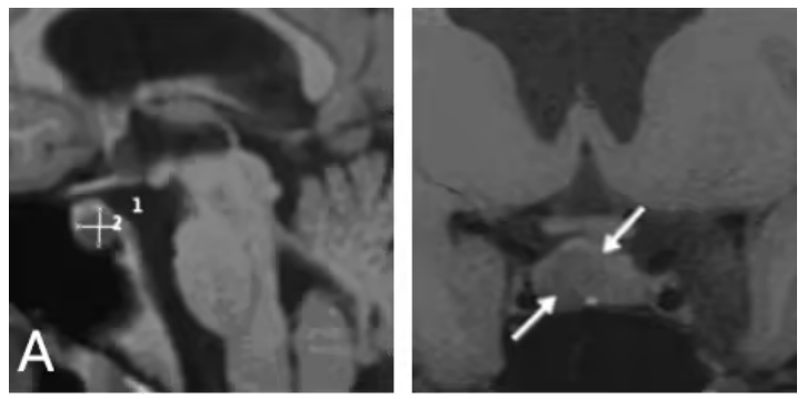
Shortly before high school graduation, Jessica developed persistent menstrual disturbance. Blood tests and MRI imaging revealed a small intrasellar pituitary tumor with leftward displacement of intact pituitary parenchyma. The patient was initiated on first-line dopamine agonist therapy (cabergoline). However, serial follow-up imaging showed progressive tumor enlargement alongside continuously rising prolactin levels, indicating definite dopamine agonist resistance.
Surgical Decision-Making
The patient’s family sought consultation with Professor Schroeder, a leading international expert in endoscopic skull base surgery affiliated with INC. The case was diagnosed as drug-resistant prolactinoma. Professor Schroeder performed endoscopic-assisted microscopic transsphenoidal tumor resection.
Postoperative Outcomes
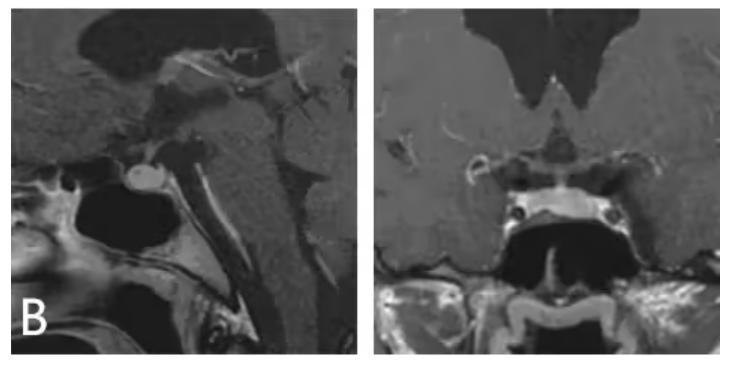
One-year postoperative MRI confirmed gross total resection of the tumor with complete recovery of displaced pituitary tissue. Endocrine reassessment demonstrated normalized serum prolactin and stable levels of other pituitary hormones, with no requirement for long-term hormone replacement therapy.
Epidemiological and Clinical Features of Pituitary Prolactinoma
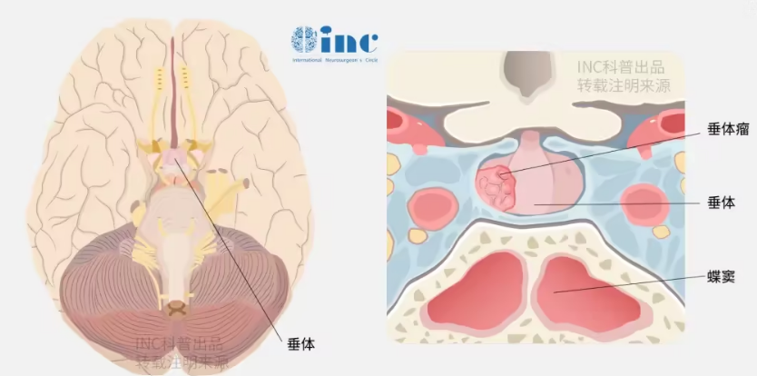
Prolactinoma, originating from pituitary lactotrophs, is the most common subtype of functional pituitary adenomas, accounting for 40% of all pituitary neoplasms. The disease presents a striking female predominance, with 80%–90% of diagnosed cases occurring in women. Female patients typically harbor small, non-invasive intrasellar tumors, predominantly manifesting with hormone-related symptoms including menstrual irregularities, galactorrhea and infertility. Mass-effect complications such as visual impairment and hypopituitarism are relatively rare in this population.
Treatment Strategies: Clinical Decision-Making Between Medical and Surgical Intervention
First-Line Medical Therapy
Dopamine agonists (e.g., bromocriptine, cabergoline) serve as the standard first-line treatment, capable of effectively reducing tumor volume even in macroadenomas. Nevertheless, approximately 10%–15% of patients develop drug resistance or intolerable adverse effects, necessitating alternative management.
Surgical Indications
As emphasized by Professor Schroeder, microsurgical resection is the preferred alternative for patients with refractory disease or drug intolerance. Transsphenoidal approaches (endoscopic and microscopic) are applicable for the majority of pituitary adenomas, particularly midline lesions with suprasellar extension limited above the internal carotid artery bifurcation.
Technical Key Points of Transsphenoidal Surgery
Advantages of the Transsphenoidal Approach
The endoscopic endonasal transsphenoidal approach provides superior panoramic visualization and flexible manipulation, facilitates protection of the superior hypophyseal artery and its branches, and has become the mainstream surgical modality for pituitary tumor resection.
Core Surgical Principles
Tumor dissection: Pituitary apoplexy often presents with hemorrhagic and necrotic changes. Tumor tissue is typically soft. After dural opening, dark necrotic hemorrhagic tissue is gently evacuated via curettes and suction devices. Over-dissection of normal pituitary parenchyma is strictly avoided to preserve endogenous endocrine function.
CSF leak prevention: Intraoperative dural injury is minimized. Any dural defect is immediately reconstructed using artificial dura, autologous fascia or fat grafts to reduce postoperative intracranial infection risk.
Neurovascular protection: For patients with preoperative oculomotor palsy caused by cavernous sinus compression, special attention is paid to protecting the internal carotid artery and cranial nerve complexes during dissection.
Individualized Treatment and Integrated Surgical Technology
Professor Schroeder advocates individualized management of pituitary adenomas based on tumor size, anatomical location and hormonal secretory characteristics. The integration of neuroendoscopic minimally invasive techniques and intraoperative neuromonitoring significantly improves gross total resection rates and long-term quality of life, achieving precise tumor eradication while maximizing preservation of pituitary function.
This clinical case — beginning with gynecological endocrine symptoms and ultimately requiring neurosurgical intervention — highlights the critical value of interdisciplinary collaboration in the standardized management of complex pituitary neuroendocrine tumors.