2026-07-03
Pituitary neuroendocrine neoplasms have long been categorized as benign lesions under traditional clinical frameworks, with standard management consisting of pharmacotherapy and surgical resection that yield satisfactory outcomes for most patients. Nevertheless, invasive pituitary neuroendocrine tumors (APT) account for approximately 10% of all pituitary neoplasms. These lesions display an extremely high recurrence propensity, exhibit invasive histological and radiological features typical of malignant tumors, and carry an inherent risk of metastatic dissemination. This report describes a 16-year-old female patient with a giant invasive pituitary neuroendocrine tumor compressing the ophthalmic artery, optic nerve and adjacent vital structures, resulting in severe visual deterioration. After failed medical therapy and recurrent disease following initial surgery, salvage surgical resection became the critical therapeutic question.
Sixteen-year-old Lina presented with right eye vision reduced to 0.1, intractable headache and menstrual irregularities. Imaging identified an intracranial pituitary prolactinoma (Figure 1) exerting mass effect on the optic nerve.
Initial Lesion
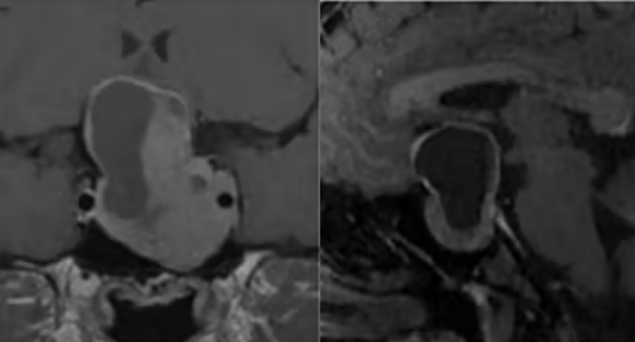
Figure 1: Baseline tumor imaging
Lina was initiated on daily cabergoline, yet no meaningful tumor regression was observed, signifying medical treatment failure. She subsequently underwent transnasal endoscopic resection, achieving partial tumor removal that relieved cerebral compression and partially restored visual function. Eight years later, Lina developed recurrent visual loss and ptosis, and was hospitalized due to spontaneous intracerebral hemorrhage. Imaging confirmed tumor recurrence, prompting a second operation; residual tumor tissue remained after the procedure.
Four months thereafter, persistent visual impairment led to her referral to neurosurgery. MRI demonstrated residual tumor, concurrent secondary empty sella syndrome, and inferior displacement of the anterior communicating artery complex within the sellar region (Figure 2).
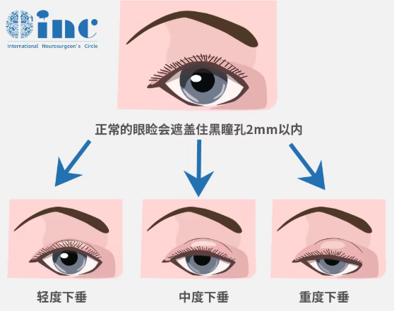
Normal eyelid position covers the black pupil by no more than 2 mm; ptosis denotes pathological drooping of the upper eyelid.
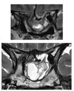
Figure: Imaging showing tumor recurrence 8 years after primary resection, complicated by intracerebral hemorrhage and sellar floor descent
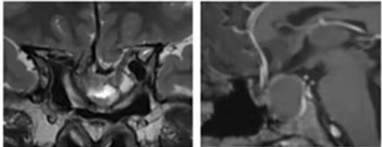
Figure 2: Preoperative MRI demonstrating residual tumor after second surgery and radiological features of secondary empty sella syndrome
The third operation was performed by Professor Florian from INC, en bloc gross total resection of the tumor was accomplished. One-year postoperative MRI confirmed complete tumor extirpation with fat graft reconstruction of the empty sella (Figure 3). Postoperatively, the patient’s left visual field improved, visual acuity stabilized at 0.3, and right eye vision rose dramatically from a preoperative 0.03 to 1.0, achieving normal visual function.
One-Year Postoperative MRI
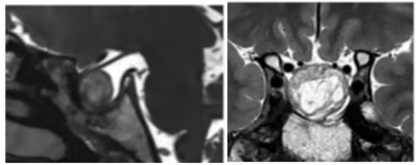
Figure 3: MRI scan one year after tertiary resection
Clinical Characterization of Invasive Pituitary Tumors
The clinical complexity of pituitary neoplasms stems not only from their anatomical location but also from a distinct subtype: lesions that continue to proliferate and invade surrounding tissues despite surgical resection, medication and radiotherapy. Clinically termed invasive pituitary tumors (APT), their core defining feature is persistent disease progression after standard multimodal therapy.
A critical distinction must be clarified prior to further discussion: residual tumor visualized on postoperative MRI is a separate entity from true tumor recurrence. In patients receiving subtotal resection, persistent postoperative tumor remnants that grow slowly do not constitute recurrence, but simply represent incompletely excised tissue. True recurrence refers to de novo regrowth of residual tissue after a period of stable disease following gross total or subtotal resection. These two scenarios dictate vastly different subsequent treatment algorithms; conflation of the two may misdirect clinical decision-making.
Accordingly, the extent of resection achieved during primary surgery forms the cornerstone of all treatment. This explains why pituitary tumor resection mandates management by specialized, experienced neurosurgeons: maximal initial resection correlates strongly with superior long-term prognosis.
Serial MRI surveillance serves as the primary imaging modality throughout the disease course, acquired preoperatively, postoperatively and at all follow-up intervals. Additional CT is indicated when bony invasion is suspected. The core objective of each follow-up scan is direct comparison with prior imaging; clinical focus lies not on absolute single-timepoint tumor size, but on volumetric change and progressive invasion of adjacent structures. Unidimensional and bidimensional diameter measurements may fail to detect subtle treatment response or disease progression, especially for multilobulated, multifocal and irregularly shaped tumors, where measurements across different imaging planes yield significant numerical discrepancies. Volumetric assessment delivers far more reliable quantitative data, yet its routine clinical adoption remains limited and merits wider implementation.
The Knosp grading system, ranging from Grade 0 to Grade 4, is the universal tool for quantifying cavernous sinus invasion, with higher grades corresponding to deeper intrasinus infiltration. This classification guides surgical difficulty stratification, residual tumor risk prediction and postoperative recurrence surveillance. Machine learning combined with radiomic analysis represents an emerging research frontier, with promising potential for non-invasive prediction of pituitary tumor proliferative activity in future clinical practice.
Imaging alone provides insufficient diagnostic information. Serial hormonal testing is mandatory, as shifts in endocrine markers often precede radiological evidence of disease progression. Tumor response to targeted pharmacotherapy also constitutes an auxiliary criterion for evaluating invasive potential.
The Knosp grading system is the most widely adopted classification for quantifying cavernous sinus invasion by pituitary macroadenomas.
Modified Knosp Grading System
This grading framework relies on coronal MRI of the cavernous sinus, drawing three vertical reference lines along the medial margin, midpoint and lateral margin of the internal carotid artery (ICA) C4 and C6 segments to characterize adenoma-cavernous sinus relationships.
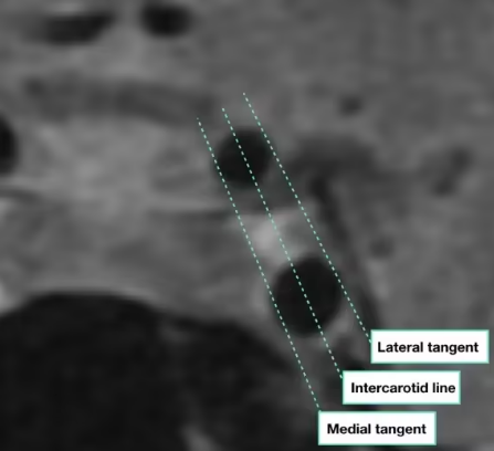
As illustrated on coronal sequences, three vertical lines are demarcated along the medial edge, central axis and lateral edge of the ICA; grading is assigned based on the adenoma’s positional relationship to these landmarks:
Grade 0: Tumor confined medial to the medial ICA tangent line
Grade 1: Adenoma extends beyond the medial tangent but does not cross the mid-carotid reference line
Grade 2: Tumor crosses the mid-carotid line without exceeding the lateral ICA tangent
Grade 3: Tumor extends lateral to the outer ICA tangent
Grade 3A: Superior extension of tumor into the cavernous sinus beyond the lateral tangent
Grade 3B: Inferior extension of tumor into the cavernous sinus beyond the lateral tangent
Grade 4: Complete encasement of the intracavernous internal carotid artery
Therapeutic Strategies for Invasive Pituitary Tumors
Prior to initiating targeted therapy for confirmed local recurrence, systemic staging with PET and craniospinal MRI is essential to screen for distant metastatic lesions.
Transnasal approaches constitute the standard corridor for pituitary surgery. Endoscopic techniques offer wider surgical exposure and direct visualization of intrasellar anatomy, facilitating more complete resection of tumors invading the cavernous sinus and parasellar compartments.
The primary clinical challenge of APT lies in high intrinsic medical treatment failure rates, alongside unrelenting mass effect on the optic nerve, cranial nerves and adjacent structures; watchful waiting inevitably worsens clinical morbidity. Reoperation carries tangible technical obstacles, including tissue fibrosis, distorted anatomical planes, and elevated risk of cranial nerve dysfunction for laterally invasive cavernous sinus disease. Nonetheless, repeat or staged surgical debulking warrants thorough discussion for all APT patients prior to initiating chemotherapy or targeted therapy.
A prevalent clinical misconception holds that surgery is futile if gross total resection cannot be achieved. This logic does not apply to APT. Even incomplete debulking alleviates compressive visual deficits and reduces overall tumor burden, enabling subsequent chemotherapy and targeted agents to act on a smaller neoplastic volume and improving therapeutic efficacy.
Conclusion
Over recent years, the pituitary neuroendocrinology field has undergone a fundamental paradigm shift: pituitary adenomas are no longer regarded as purely benign endocrine disorders, but reclassified as neuroendocrine neoplasms with malignant transformation potential. APT is now positioned along a continuous spectrum of malignant potential, as an intermediate entity between standard-risk pituitary adenomas and pituitary carcinoma. This revised framework compels pituitary specialists to adopt comprehensive neuro-oncologic management algorithms. Central to this model is iterative discussion of resectability at every disease stage, as surgical intervention optimizes symptomatic control and potentiates the efficacy of adjuvant systemic therapies.
This shift also raises unresolved clinical questions, including optimal timing for systemic intervention (earlier introduction of temozolomide [TMZ], appropriate treatment discontinuation criteria, applicability of the Stupp protocol to selected APT cases), and the emerging role of experimental oncologic therapies adapted from systemic tumor management, such as targeted molecular agents and immunotherapy. Ultimately, regardless of whether lesions are labeled pituitary adenomas or pituitary neuroendocrine tumors, neurosurgical expertise remains indispensable at all disease stages, requiring clinicians to adapt to evolving pathological classifications and expanding therapeutic armamentaria.








